Κατέβασμα παρουσίασης
Η παρουσίαση φορτώνεται. Παρακαλείστε να περιμένετε

1
Κακώσεις –Παθήσεις –Μηρού-Γόνατος
Ανατομία dsfsf Κακώσεις –Παθήσεις –Μηρού-Γόνατος dsfsf dsfsf Ελευθερία Θωμαΐδου, Pt

2
Some common bony landmarks
Adductor tubercle Medial and lateral femoral epicondyles and condyles Intercondylar fossa Medial and lateral tibial condyles Medial and lateral tibial plateau Intercondylar notch Intercondylar eminence Tibial tuberosity Patella Head of fibula Please note these are not the only bony landmarks

3
Prime Movers of the Knee
Extensors (Quads) Rectus Femoris Vastus Medialis Vastus Lateralis Vastus Intermedius Primary Flexors (Hams) Semimembranosus Semitendinosus Biceps Femoris Secondary Flexors Sartorius Gracilis Gastrocnemius Medial Rotators Semitendinosus Semimembranosus Lateral Rotator Biceps femoris
 Rectus Femoris. Vastus Medialis. Vastus Lateralis. Vastus Intermedius. Primary Flexors (Hams) Semimembranosus. Semitendinosus. Biceps Femoris. Secondary Flexors. Sartorius. Gracilis. Gastrocnemius. Medial Rotators. Semitendinosus. Semimembranosus. Lateral Rotator. Biceps femoris.")
4
ΚαταγΜατα Διαφυσησ Μηριαιου οστου
ΚαταγΜατα Διαφυσησ Μηριαιου οστου Συμβαίνουν σε όλες τις ηλικίες, συχνότερα σε νέα άτομα ύστερα από τροχαία ατυχήματα, πτώσεις από μεγάλα ύψη. Συμβαίνουν έπειτα από δράση ισχυρής βίας-> ρήξη μυών ,αγγείων, μαλακών μορίων-> κίνδυνος για την ακεραιότητα του μέλους. Επηρεάζεται η αιμάτωση του μηριαίου οστού( αγγεία του περιόστεου, ενδομυελικά αγγεία)
")
5
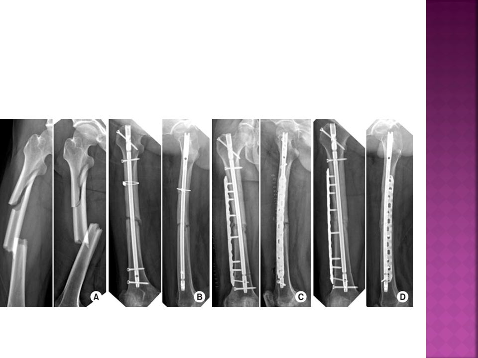
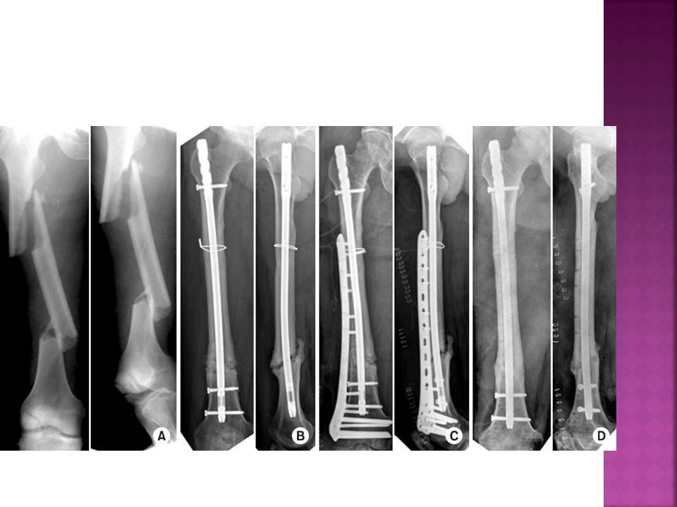
Ταξινομηση καταγματων διαφυσης
Ταξινομούνται κατά Winquist ανάλογα με το βαθμό συντριπτικότητας:

6
Τύπος Ο: Καμία παρεκτόπιση
Τύπος Ι: Λοξά κατάγματα με ελάχιστο διαχωρισμό των τεμαχίων Τύπος ΙΙ: Ύπαρξη οστικού τεμαχίου( παρασχίδα) Τύπος ΙΙΙ: Οι διαστάσεις του οστικού τεμαχίου κυμαίνονται από 50%-100% του πάχους της διάφυσης
 Τύπος ΙΙΙ: Οι διαστάσεις του οστικού τεμαχίου κυμαίνονται από 50%-100% του πάχους της διάφυσης.")
9
Καταγματα του κατω περατος του μηριαου οστου
Βαριές κακώσεις που επηρεάζουν την κινητικότητα του γόνατος Σε νέα άτομα προκαλούνται από τη δράση βίας, σε τροχαία ατυχήματα Σε ηλικιωμένους λόγω οστεοπόρωσης από την πτώση

10
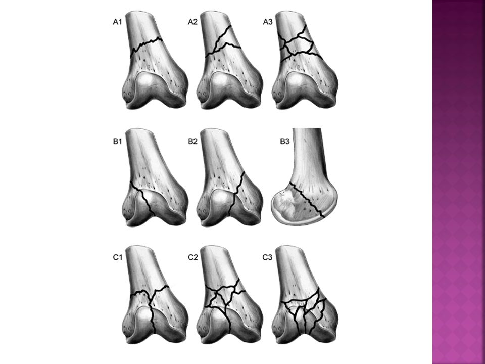
Αντιμετώπιση Ταξινομούνται κατά την ΑΟ σε:
Α. Εξωαρθρικά κατάγματα -> υπεκονδύλια Β. Ενδοαρθρικά( ενός κονδύλου) C. Συνδυασμός εξωαρθρικού με ενδοαρθρικό-> τύπου Τα ή Υ Στα παραπάνω υπάρχουν και υποκατηγορίες ανάλογα με τη βαρύτητα Κλινική εικόνα Διόγκωση και παραμόρφωση του γόνατος Περιορισμός ή και κατάργηση της κινητικότητας της άρθρωσης λόγω πόνου Αντιμετώπιση Συντηρητική : σπάνια με σκελετική έλξη ή ΜΚ γύψου Χειρουργική :Γωνιώδης ήλος-πλάκα, πλάκα με βίδα συμπίεσης, ενδομυελικοί ήλοι
 C. Συνδυασμός εξωαρθρικού με ενδοαρθρικό-> τύπου Τα ή Υ. Στα παραπάνω υπάρχουν και υποκατηγορίες ανάλογα με τη βαρύτητα. Κλινική εικόνα. Διόγκωση και παραμόρφωση του γόνατος. Περιορισμός ή και κατάργηση της κινητικότητας της άρθρωσης λόγω πόνου. Αντιμετώπιση. Συντηρητική : σπάνια με σκελετική έλξη ή ΜΚ γύψου. Χειρουργική :Γωνιώδης ήλος-πλάκα, πλάκα με βίδα συμπίεσης, ενδομυελικοί ήλοι.")
13
Καταγμα επιγονατιδασ Αποτελεί το 1% των καταγμάτων του σκελετού
Μηχανισμός κάκωσης α)Άμεση κάκωση όπως πτώση πάνω στην επιγονατίδα ή πλήξη β) Έμμεση βία λόγω της σύσπασης του τετρακεφάλου μυός Κλινική εικόνα Πόνος αυτόματος και με τη ψηλάφηση ή πίεση, διόγκωση του γόνατος( αίμαρθρο), αδυναμία έκτασης του γόνατος
Άμεση κάκωση όπως πτώση πάνω στην επιγονατίδα ή πλήξη. β) Έμμεση βία λόγω της σύσπασης του τετρακεφάλου μυός. Κλινική εικόνα. Πόνος αυτόματος και με τη ψηλάφηση ή πίεση, διόγκωση του γόνατος( αίμαρθρο), αδυναμία έκτασης του γόνατος.")
14
Patella Fractures

15
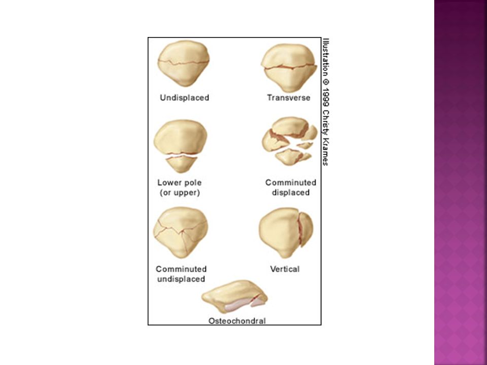
Patella Fractures Several primary types of patellar fractures have been identified, each with separate diagnostic, imaging, and management considerations. The primary types include transverse, vertical, marginal, and osteochondral fractures.

16
Ταξινόμηση Ρωγμώδη Εγκάρσια με διάσταση( πιο συχνά) Άνω η κάτω πόλου της επιγονατίδας Επιμήκη( κάθετα) του πλάγιου χείλους της Συντριπτικά Οστεοχόνδρινα( σπάνια) σε νέα άτομα ετών
 σε νέα άτομα ετών.")
18
Οστεοσύνθεση με βελόνες Kirschner
και συρμάτινη αγκύλη ταινία ελκυσμού( tension bad)
")
19
Ρηξη τενοντα 4φαλου-επιγονατιδικου συνδεσμου
Ρηξη τενοντα 4φαλου-επιγονατιδικου συνδεσμου Προκαλείται κατάργηση του εκτατικού μηχανισμού

20
Εξαρθρημα της επιγονατιδας
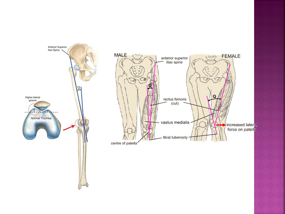
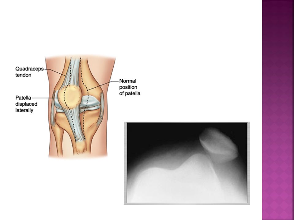
Είναι σχεδόν πάντα πλάγιο έξω Διακρίνεται σε : Α. οξύ τραυματικό (τραυματισμός με το γόνατο σε κάμψη) Β. Σε υποτροπιάζον( καθ’ έξιν)οφείλεται σε συγγενείς ανωμαλίες ή και επίκτητες Αύξηση της γωνίας Q Δυσπλαστική επιγονατίδα Υποπλασία έξω μηριαίου κονδύλου Συνδεσμική χαλάρωση Ατροφία του έσω πλατέος μυός ή υπερτροφία του έξω
 Β. Σε υποτροπιάζον( καθ’ έξιν)οφείλεται σε συγγενείς ανωμαλίες ή και επίκτητες. Αύξηση της γωνίας Q. Δυσπλαστική επιγονατίδα. Υποπλασία έξω μηριαίου κονδύλου. Συνδεσμική χαλάρωση. Ατροφία του έσω πλατέος μυός ή υπερτροφία του έξω.")
23
Δοκιμασία φόβου επικείμενης εξάρθρωσης επιγονατίδας

24
The Knee Largest synovial joint in body and it also the most complex
Simply put it is a hinge joint However more technically it is a mobile trocho- ginglymus (Pivotal hinge joint) since it not only allows flexion and extension but also slight medial and lateral rotation Since in humans the knee supports nearly the whole weight of the body, it is the joint most vulnerable both to acute injury and the development of osteoarthritis Osteoarthritis Osteoarthritis , is a group of diseases and mechanical abnormalities involving degradation of joints, including articular cartilage and the subchondral bone next to it... .
 since it not only allows flexion and extension but also slight medial and lateral rotation. Since in humans the knee supports nearly the whole weight of the body, it is the joint most vulnerable both to acute injury and the development of osteoarthritis Osteoarthritis. Osteoarthritis , is a group of diseases and mechanical abnormalities involving degradation of joints, including articular cartilage and the subchondral bone next to it... .")
25
Εξαρθρημα του ΓΟνατος Συμβαίνει σπάνια, λόγω των ισχυρών συνδέσμων και μυών Παρεκτόπιση του ενός οστού σε σχέση με το άλλο ( πρόσθιο – οπίσθιο- πλάγιο) Σοβαρότατη κάκωση. Ιδιαίτερα το οπίσθιο καθώς θέτει σε κίνδυνο τα αγγεία και νεύρα της ιγνυακής κοιλότητας
 Σοβαρότατη κάκωση. Ιδιαίτερα το οπίσθιο καθώς θέτει σε κίνδυνο τα αγγεία και νεύρα της ιγνυακής κοιλότητας.")
27
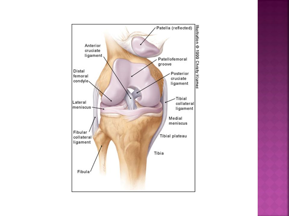
Κακωσεις συνδεσμων γονατος
Ρήξη έσω θυλακοσυνδεσμικού συστήματος( ΕΠΣ) Νόσος των Pellegrini-Stieda Ρήξη του έξω θυλακοσυνδεσμικού συστήματος (ΕξΠΣ) Ρήξη του Πρόσθιου Χιαστού Συνδέσμου ACL Ρήξη του οπίσθιου Χιαστού Συνδέσμου PCL
 Νόσος των Pellegrini-Stieda. Ρήξη του έξω θυλακοσυνδεσμικού συστήματος (ΕξΠΣ) Ρήξη του Πρόσθιου Χιαστού Συνδέσμου ACL. Ρήξη του οπίσθιου Χιαστού Συνδέσμου PCL.")
28
Cross Section

29
Ρηξη εσω πλαγιου συνδεσμου
Μηχανισμός κάκωσης Βία που δρα στην εξωτερική επιφάνεια του γόνατος και οδηγεί σε απαγωγή της κνήμης( δύναμη βλαισότητας) ενώ το γόνατο βρίσκεται σε έκταση ή μικρή κάμψη 1ου- 2ου 3ου βαθμού Κλινική εικόνα Διόγκωση, ευαισθησία, περιορισμός κίνησης, πόνος δυσκολία στη βάδιση
 ενώ το γόνατο. βρίσκεται σε έκταση ή μικρή κάμψη. 1ου- 2ου 3ου βαθμού. Κλινική εικόνα. Διόγκωση, ευαισθησία, περιορισμός κίνησης, πόνος δυσκολία στη βάδιση.")
30
Δοκιμασία βλαισής φόρτισης

31
Ρηξη του εξω πλαγιου συνδεσμου
Συμβαίνει σπάνια Μηχανισμός κάκωσης Βίαιη προσαγωγή της κνήμη λόγω χτυπήματος στην έξω επιφάνεια Τραυματίζονται ταυτόχρονα η ITB και ο τένοντας του ιγνυακού

32
Δοκιμασία ραιβής φόρτισης

33
νΟσος των Pellegrini- stieda
Πρόκειται για οστεοποίηση της έκφυσης του έσω πλαγίου συνδέσμου

34
Ρηξη προσθιου χιαστου συνδεσμου
Συχνός αθλητικός τραυματισμός Όταν βίαιη δύναμη ωθήσει την κνήμη προς τα μπρος σε σχέση με το μηρό Συχνά η κάκωση συνδυάζεται και με ρήξη έσω μηνίσκου και έσω πλαγίου συνδέσμου ( ατυχής τριάδα του O’ Donohue) Αίμαρθρο
 Αίμαρθρο.")
35
Δοκιμασία Lachman- Νούλη(1875)
Δοκιμασία πρόσθιου συρταρωτού Τεστ θετικό όταν η μετατόπιση είναι > 5mm SOSss στην πρώτη εξέταση

37
Arthroscopy

38
Ρηξη οπισθιου χιαστου συνδεσμου
Συχνότητα 1:10 σε σχέση με τον ΠΧΣ Πτώση από ύψος με στήριξη στο γόνατο που βρίσκεται σε κάμψη Βίαιη ώθηση της κνήμης προς τα πίσω σε σχέση με το μηριαίο Αίμαρθρο

39
Δοκιμασία οπίσθιου συρταρωτού
sossss

40
Κακωσεις μηνισκων γονατος
Ρήξη έσω μηνίσκου Συχνότερα σε αναλογία 1:2 σε σχέση με τον έξω Ρήξη έξω μηνίσκου

41
Δοκιμασία Αpley test

42
Δοκιμασία Mc Murray

43
Συνδρομο της υμενικης πτυχης( plica syndrome)
Σπάνιο αίτιο πόνου(2%)στην άρθρωση του γόνατος Η έσω επιγονατιδική πτυχή παρουσιάζει φλεγμονή και να προκαλεί πόνο στην ΕΜ άρθρωση λόγω προστριβής στην επιγονατίδα ή στον μηριαίο κόνδυλο Η επιγονατίδα κινείται ανώμαλα ή αναπηδά κατά την κάμψη
στην άρθρωση του γόνατος. Η έσω επιγονατιδική πτυχή παρουσιάζει φλεγμονή και να προκαλεί πόνο στην ΕΜ άρθρωση λόγω προστριβής στην επιγονατίδα ή στον μηριαίο κόνδυλο. Η επιγονατίδα κινείται ανώμαλα ή αναπηδά κατά την κάμψη.")
44
Ιγνυακη κυστη( κυστη βακεr)
Προβολή του αρθρικού υμένα και συσσώρευση του αρθρικού υγρού μέσα στον ιγνυακό βόθρο η συσσώρευση του υγρού μπορεί να οφείλεται και στου τενόντιους θύλακες των οπίσθιων μηριαίων μυών Περιορίζει την κάμψη του γόνατος

45
Γόνατο του Αλτη( jumper’s knee)
Τενοντίτιδα του επιγονατιδικού τένοντα Λόγω καταπόνησης, συμβαίνουν μικροτραυματισμοί και φλεγμονή στον καταφυτικό τένοντα του 4 φάλου και τον επιγονατιδικό Πόνος κατά την παθητική διάταση του τένοντα και την υπο αντίσταση έκταση του γόνατος

46
Συνδρομο τριβησ λαγονοκνημιαιας ταινιας
Κάκωση υπέρχρησης σε δρομείς και ποδηλάτες Οφείλεται στην τριβή της ταινίας πάνω στον έξω μηριαίο υπερκονδύλιο κύρτωμα Στις 30ο περίπου το μεταβατικό όριο Η ανελαστικότητα της ταινίας(ober test), προπονητικά σφάλματα, τρέξιμο σε κατηφόρα είναι προδιαθεσιακοί παράγοντες
, προπονητικά σφάλματα, τρέξιμο σε κατηφόρα είναι προδιαθεσιακοί παράγοντες.")
47
Δoκιμασία συμπίεσης Noble

48
Should pay attention to the tibial plateau.
If you draw a line from the lateral border of the femoral condyle to the lateral border of the tibial condyle. This should be perpendicular and have no more than 5mm of adjacent tibia next to it = LATERAL TIBIAL LINE Check the adequacy of the medial and lateral joints. Is there narrowing? There is no evidence of a bony injury or abnormality seen on the images provided.

49
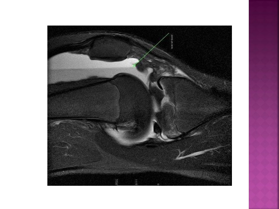
Effusion or lipohaemoarthrosis
Check the soft tissue Effusion or lipohaemoarthrosis Check Alignment tibial plateau aligned with femoral condyle. Check bone-cortex, trabecular pattern, check joint space Flabella- norm variant Bipartite patella- norm variant No evidence of a bony injury or abnormality seen on the lateral view provided

50
Knee Trauma 5% of all physeal fractures at distal femur.
Common mechanism is hyperextension causing anterior displacement of epiphysis. Salter Harris Type I injury. Leg length discrepancy of > 2 cm may develop in 1/3 of pts.

51
Trauma Knee

52
Παρεση περονιαιου νευρου
Το κοινό περονιαίο νεύρο είναι ευάλωτο στο σημείο που τυλίγεται γύρω από την περόνη Μπορεί να τραυματιστεί από άμεση πλήξη, έντονη κρυοθεραπεία, ή εφελκυσμό Πόνος, αιμωδία, καυσαλγία, αδυναμία ραχιαίων καμπτήρων του ακρου ποδός, πτώση πέλματος

53
Trauma Knee (Cont.)
")
54
Effusion NORMAL= There is NO increase in the distance
< 5mm NORMAL= There is NO increase in the distance Between pre-femoral and suprapatellar bursa. > 5MM ABNORMAL= Distension of suprapatellar bursa. Distance between prefemoral and suprafemoral bursa is > 5mm Also watch for an increase space posterior to the quadriceps tendon. A large joint effusion may cause you not to see this at all on the lateral view. Be aware that effusions can be caused by other processes apart form fractures, ligamentous injury, infection or arthritis.

55
Lipohaemarthrosis If you see a Lipohaemoarthrosis then this is indicative of an intra-articular fracture. This is due to marrow being released into the joint. The fat sits above a layer of fluid (normally blood). Therefore you are seeing a fat-fluid level. Liophaemoartrosis detected then send for an orthopaedic referral. It is the common sign associated with a tibial plateau fracture.
. Therefore you are seeing a fat-fluid level. Liophaemoartrosis detected then send for an orthopaedic referral. It is the common sign associated with a tibial plateau fracture.")
57
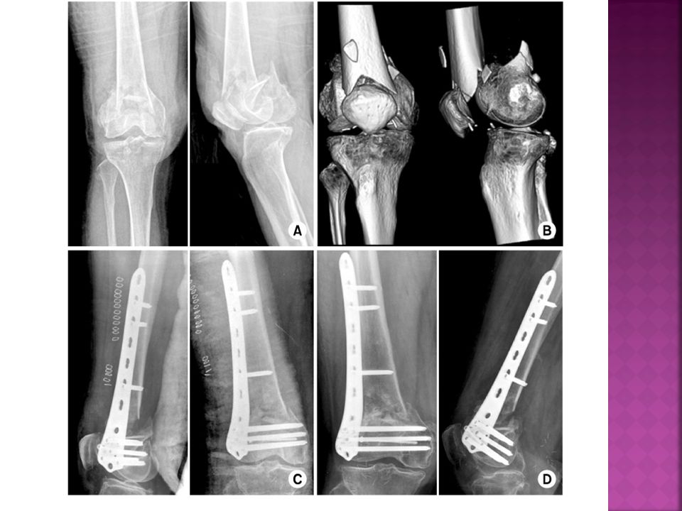
Καταγματα του ανω ακρου της κνημησ( κνημιαιου plateau)
Πρόκειται για ενδοαρθρικά κατάγματα Κάταγμα του έξω μηριαίου κονδύλου>>έσω Μηχανισμός κάκωσης Αποτέλεσμα πλάγιας κάμψης και κατακόρυφης συμπίεσης Συνοδεύεται και από συνδεσμική ρήξη

58
Ταξινόμηση Τύπος Ι. Σφηνοειδή διαχωριστικά του έξω κονδύλου
Τύπος ΙΙ. Συμπιεστικά του έξω κονδύλου με καθίζηση Τύπος ΙΙΙ. Με κεντρική καθίζηση της αρθρικής επιφάνειας Τύπος ΙV. Κάταγμα του έσω κνημιαίου κονδύλου Τύπος V. Κάταγμα και των δύο κονδύλων με μορφή Τ ή Υ Τύπος VI. Κάταγμα plateau οποιασδήποτε μορφής με ταυτόχρονο μεταφυσιακό κάταγμα ( συντριπτικό)
")
59
Normal Tibial Plateau L Medial tibial plateau Lateral tibial plateau
On the lateral- tibial plateau should align with femoral condyles. Anterior displacement –if tibial plateau shifts forward suspect rupture of anterior cruciate ligament. Posterior displacement can occur in a rupture of the posterior cruciate ligament. Look at the bone- is there an increase in sclerosis seen within the proximal area of the tibia if there is this may be a subtle tibial plateau fracture. Step defect may also be the only sign of a subtle injury. Look at the integrity of the joint space, widening may indicate a tibial plateau fracture L Medial tibial plateau Lateral tibial plateau Tibial Plateau

60
Lateral Tibial Plateau
There is an intra-articular fracture of the lateral tibial plateau of the right knee seen on the views provided. A lipohaemarthrosis is not demonstrated as a horizontal beam lateral radiograph has not been undertaken.

61
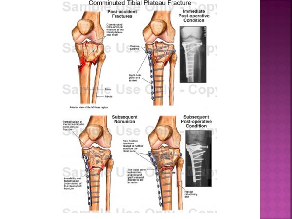
Tibial Plateau fracture
Most often in women > 50 years Mechanism of injury: twisting injury, bumper striking the knee Valgus force causes the femoral condyle to impact on the plateau 70-80% cases involve lateral tibial plateau Degree of the depression of the fracture determines if surgery is required. Normally go for CT to assess. Orthopaedic intervention normally involves tibial plate and screws.

62
Medial Tibial Plateau Note the step in the cortex and the increase in sclerosis These are less frequent than fractures involving the lateral condyle. Mechanism: forced impact from the medial femoral condyle onto the tibial plateau There is an intra-articular fracture of medial tibial plateau of the left knee present on the AP view provided. Please note, a lateral view has not been undertaken. L

63
Tibial Plateau Trauma

65
Tibial stress fractures
Subtle Common in runners and dancers Look for an presence of a sclerotic band or a periosteal reaction/cortical thickening What is the differential for periosteal reaction? There is a sclerotic band seen within the proximal third of the Rt.tibia with slight periosteal reaction noted. Its appearance is consistent with a stress fracture at this level. R

67
Avulsion fracture- lateral tibial condyle
Could be known as a Segond fracture. Always adjacent to lateral tibial plateau just below articular surface Associated injury to anterior cruciate ligament/meniscus There is an avulsion fracture seen off the lateral tibial condyle just inferior to the lateral tibial plateau. This does not appear to involve the articular surface. Please note, only an AP view of the knee has been provided.

68
Segond Lesion An avulsion fracture of the lateral tibial plateau, also known as a Segond fracture, predicts the presence of an ACL rupture in 75% to 100% of cases.

69
Segond Lesion

70
Osgood Schlatters Μικροτραυματισμοί από τον επαναλαμβανόμενο εφελκυσμό του επιγονατιδικού συνδέσμου στο κνημιαίο κύρτωμα Κατά την εφηβεία Συνήθως αγόρια με δραστηριότητα άλματα ή δρομικά Μπορεί να είναι άμφω(25-50%) Πόνος και οίδημα στο κνημιαίο κύρτωμα Soft tissue swelling and compatible history assist diagnosis
 Πόνος και οίδημα στο κνημιαίο κύρτωμα. Soft tissue swelling and compatible. history assist diagnosis.")
72
OA and the knee joint

73
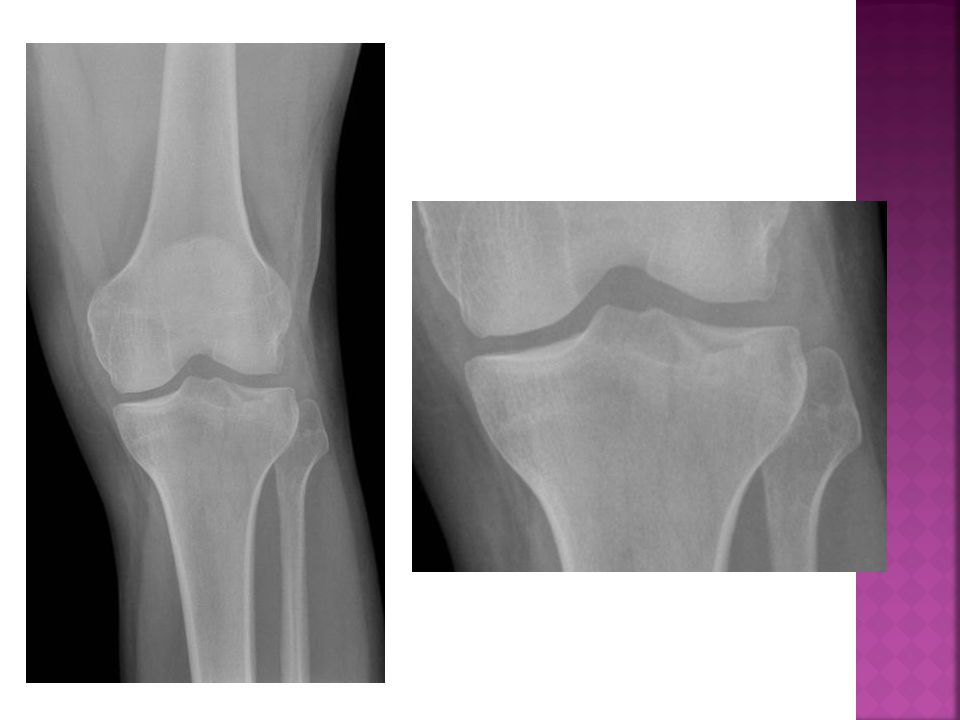
Οστεοαρθριτιδα γονατος
Στένωση του μεσάρθριου διαστήματος Σχηματισμός οστεοφύτων Παρουσία κυστών Συνήθως επηρεάζεται πρώτα το έσω διαμέρισμα There is joint space narrowing of the medial joint compartment seen with sub articular sclerosis noted. With osteophyte formation noted on the medial tibial condyle. Its appearance is characteristic of osteoarthritis R

74
OA Knee Joint space narrowing Osteophyte formation
Subchondral sclerosis Cyst formation Varus Deformity

75
Advanced OA changes Effacement of medial joint space.
Sub-chondral cysts. Multiple Osteophytes. Sclerotic articular surfaces. Varus Deformity (Bow legged) If this is confusing, just remember that "varus = inward" and "valgus = outward" and always refers to the direction that the distal part of the joint points. The English-speaking orthopaedic’s mnemonic is: valgus with a g as in gum means knees that stick together, and varus means a variance or divergence.
 If this is confusing, just remember that varus = inward and valgus = outward and always refers to the direction that the distal part of the joint points. The English-speaking orthopaedic’s mnemonic is: valgus with a g as in gum means knees that stick together, and varus means a variance or divergence.")
76
Valgus Deformity Femur
More common in females. In valgus knee, ligament balancing is more difficult to fix. Knee Joint Tib and Fib

77
Radiographically Medial joint space narrowing occurs first.
Can lead to uni- compartment knee replacement. Osteophyte formation at TIES, lateral tibial plateau borders, and on the most lateral aspects of the femoral condyles, with some sub-chondral cyst formation.

79
Chondrocalcinosis Associated with the condition known as CPPD (Calcium pyrophosphate deposition disease) Calcification of the fibrocartilage or hyaline cartilage

80
Osteochondritis Dessicans
Osteochondritis dissecans (OCD), by definition, is a disorder of one or more ossification centres, characterized by sequential degeneration or aseptic necrosis and re-calcification. OCD lesions involve both bone and cartilage. These lesions differ from acute traumatic osteochondral fractures; however, they may manifest in a similar fashion. OCD lesions also must be differentiated from meniscal pathology. OCD causes 50% of loose bodies in the knee. The aetiology of these lesions is multi-factorial, including trauma, ischemia, abnormal ossification centres, genetic predisposition, or some combination of these factors. Little agreement exists among researchers regarding the aetiology of OCD.
, by definition, is a disorder of one or more ossification centres, characterized by sequential degeneration or aseptic necrosis and re-calcification. OCD lesions involve both bone and cartilage. These lesions differ from acute traumatic osteochondral fractures; however, they may manifest in a similar fashion. OCD lesions also must be differentiated from meniscal pathology. OCD causes 50% of loose bodies in the knee. The aetiology of these lesions is multi-factorial, including trauma, ischemia, abnormal ossification centres, genetic predisposition, or some combination of these factors. Little agreement exists among researchers regarding the aetiology of OCD.")
81
Tumours of the Distal Femur
- Non-ossifying fibroma - Osteosarcoma - Giant Cell Tumour of Bone - Ewing's sarcoma - Osteochondroma - Chondroblastoma - Chondromyxoid fibroma - Eosinophilic granuloma - Aneurysmal bone cyst - Giant cell sarcoma - Fibrous Histiocytoma - Fibrosarcoma of bone - Desmoplastic fibroma - Gaucher's Disease

82
Tumours of the Proximal Tibia
- Non-ossifying Fibroma Osteochondroma - Osteosarcoma - Giant Cell Tumour of Bone - Ewing's sarcoma: (diaphyseal region); - Chondroblastoma - Chondromyxoid fibroma - Fibrous Dysplasia - Eosinophilic granuloma - Aneurysmal bone cyst - Simple bone cyst - Giant cell sarcoma - Fibrous Histiocytoma - Fibrosarcoma of bone; - Adamantinoma - Osteoma: middle 1/3 (rare)
; - Chondroblastoma - Chondromyxoid fibroma - Fibrous Dysplasia - Eosinophilic granuloma - Aneurysmal bone cyst - Simple bone cyst - Giant cell sarcoma - Fibrous Histiocytoma - Fibrosarcoma of bone; - Adamantinoma - Osteoma: middle 1/3 (rare)")
83
Osteosarcoma RT R

84
Possible Sources Use a start point!

Παρόμοιες παρουσιάσεις


>")


>")


