Κατέβασμα παρουσίασης
Η παρουσίαση φορτώνεται. Παρακαλείστε να περιμένετε

1
Ανατομία και Βιομηχανική
dsfsf Αγκώνας dsfsf dsfsf Ελευθερία Θωμαΐδου, Pt

2
Μαθησιακά αποτελέσματα
Κατανόηση βασικών στοιχείων ανατομίας και βιομηχανικής του αγκώνα Οστικά σημεία Αρθρώσεις Μύες Σύνδεσμοι Κατανόηση βασικών στοιχείων παθο-ανατομίας Must understand proximal, distal, medial, lateral, plantar, dorsal structure and function of bones, joints, tendons, ligaments and muscles.

3
Γενικά χαρακτηριστικά
Οι αρθρώσεις και μύες της περιοχής του αγκώνα παρέχουν : κινητικότητα και λειτουργικότητα στο άνω άκρο και την άκρα χείρα Σταθερότητα για επιδέξιες ή /και βίαιες κινήσεις του χεριού. The joints and muscles of the elbow complex are primarily designed to serve the hand. Along with the shoulder and the wrist, the elbow provides the necessary mobility and functionality to the hand. The elbow is considered crucial for providing stability for skilled and forceful movements of the hand. Many of the 15 muscles that cross the elbow provide movement at the shoulder or the wrist. The elbow joints are formed by the distal end of the humerus and the proximal ends of the radius and ulna. The inferior radioulnar joint is considered to be part of the elbow complex because it acts in conjunction with the superior radioulnar joint. ΙST/UH NMS2 3

4
Οστά Αγκώνα (πρόσθια όψη)
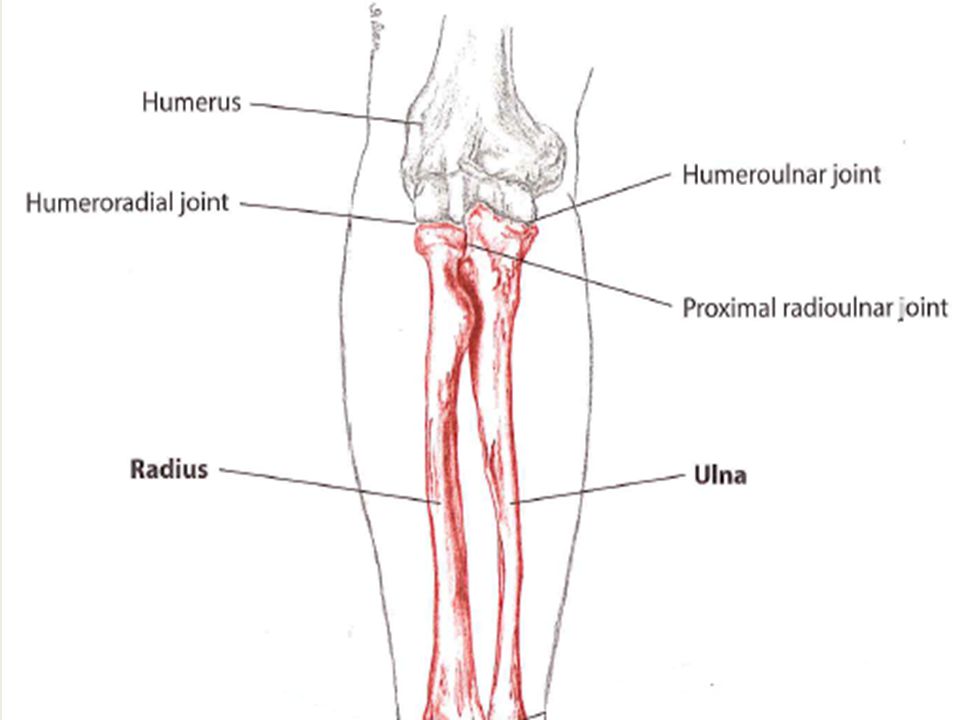
Κορωνοειδής βόθρος Κερκιδικό βοθρίο τροχιλία κόνδυλος Τροχιλιακή εντομή Κορωνοειδής απόφυση Κεφαλή κερκίδας Ωλένιο τράχυσμα (φύμα) Αυχένας κερκίδας This is an anterior view of the bony structures of the elbow complex. Distally the humerus articulates with the radius at the capitulum on the lateral side forming the humeroradial joint and the humerus articulates with the ulna at the trochlea on the medial side forming the hueroulnar joint. The concave articulating surface of the proximal ulna is known as the trochlea notch. The head of the radius also has a concave shape which allows it to articulate with the capitulum of the humerus. The articulating surfaces are covered with hyaline cartilage The cylindrical circumference of the radius head articulates with the annular ligament and radial notch of the ulna. In extension there is no contact between the radius head and the humerus. At the limit of flexion, the raidus head will move into a fossa on the humerus called the radial fossa and the coronoid process which is an anterior bony prominence on the proximal ulan will move into a fossa on the humerus called the coranoid fossa. Just distal to the coronoid process is the ulnar tuberosity from which the oblique ligament arises and attaches to the radius. This proximal forearm ligament helps the interosseous membrane in stabilizing the relationship of the radius and ulna during supination and pronation. The radius has a narrow neck just distal to the head. 4
 Αυχένας κερκίδας. This is an anterior view of the bony structures of the elbow complex. Distally the humerus articulates with the radius at the capitulum on the lateral side forming the humeroradial joint and. the humerus articulates with the ulna at the trochlea on the medial side forming the hueroulnar joint. The concave articulating surface of the proximal ulna is known as the trochlea notch. The head of the radius also has a concave shape which allows it to articulate with the capitulum of the humerus. The articulating surfaces are covered with hyaline cartilage. The cylindrical circumference of the radius head articulates with the annular ligament and radial notch of the ulna. In extension there is no contact between the radius head and the humerus. At the limit of flexion, the raidus head will move into a fossa on the humerus called the radial fossa and the coronoid process which is an anterior bony prominence on the proximal ulan will move into a fossa on the humerus called the coranoid fossa. Just distal to the coronoid process is the ulnar tuberosity from which the oblique ligament arises and attaches to the radius. This proximal forearm ligament helps the interosseous membrane in stabilizing the relationship of the radius and ulna during supination and pronation. The radius has a narrow neck just distal to the head. 4.")
5
Οστά Αγκώνα(έξω πλάγια όψη)
Οστά Αγκώνα(έξω πλάγια όψη) Υπερπαρακονδύλια ακρολοφία Παρακονδύλια απόφυση Ωλέκρανο Κερκιδική εντομή Ακρολοφία του υπτιαστή Μεσόστεο χείλος A lateral view of the bony structures of the elbow complex shows the very prominent lateral epicondyle of the humerus which can be palpated. The lateral epicondyle is the common attachment for the extensor muscles of the wrist and hand which include the extensor carpi radialis brevis, the extensor digitorum, extensor digiti minimi, and extensor carpi ulnaris. This tendinous attachment is termed the common extensor tendon. Pain in this area related to this common tendon is known as lateral epicondylalgia which will be discussed later. Proximal to the lateral epicondyle a prominent ridge on the lateral border of the humerus called the supracondylar ridge. The distal third of this ridge provides an attachment point for the extensor carpi radialis longus muscle and the upper 2/3rds of the ridge is where the brachioradialis muscle originates from. The olecranon process is the proximal most aspect of the ulna and it is the bony prominence that can be palpated posterior to the elbow joint. The triceps brachii muscle attaches to the tip of the olecranon process and via an aponeurosis to the fascia of the posterior aspect of the elbow and forearm. The radial notch is shallow concave articular surface which lies on the upper lateral aspect of the coronoid process, separated from the trochlear notch by a bony ridge. It is lined by hyaline cartilage and articulates with the side of the cylindrical head of radius. The head of the radius is held in place by the annular ligament, attached in front to the anterior margin of the radial notch and behind to a ridge just behind the notch. As mentioned before, this articulation is called the superior radioulnar joint. The lateral border of the proximal ulnar is characterised by a sharp ridge called the supinator ridge. As its name suggests the supinator ridge is the proximal attachment for the supinator muscle. It is also a very important attachment for the interosseus membrane which is formed by two layers of fibers running between the radius and ulna. The two layers of fibres run in opposite directions obliquely between the two bones and this configuration helps to strengthen the attachment between the radius and the ulna. The interosseus membrane attaches onto the ulna more distally along its medial border which is known as the interosseus border. The membrane fulfils a number of functions… It provides attachment for the deep muscles of the forearm. It is essential in maintaining the length relationship between the radius and ulna and stabilizing the forearm and the distal radioulnar joint during supination and pronation. ΙST/UH NMS2 5
 Υπερπαρακονδύλια ακρολοφία. Παρακονδύλια απόφυση. Ωλέκρανο. Κερκιδική εντομή. Ακρολοφία του υπτιαστή. Μεσόστεο χείλος. A lateral view of the bony structures of the elbow complex shows the very prominent lateral epicondyle of the humerus which can be palpated. The lateral epicondyle is the common attachment for the extensor muscles of the wrist and hand which include the extensor carpi radialis brevis, the extensor digitorum, extensor digiti minimi, and extensor carpi ulnaris. This tendinous attachment is termed the common extensor tendon. Pain in this area related to this common tendon is known as lateral epicondylalgia which will be discussed later. Proximal to the lateral epicondyle a prominent ridge on the lateral border of the humerus called the supracondylar ridge. The distal third of this ridge provides an attachment point for the extensor carpi radialis longus muscle and the upper 2/3rds of the ridge is where the brachioradialis muscle originates from. The olecranon process is the proximal most aspect of the ulna and it is the bony prominence that can be palpated posterior to the elbow joint. The triceps brachii muscle attaches to the tip of the olecranon process and via an aponeurosis to the fascia of the posterior aspect of the elbow and forearm. The radial notch is shallow concave articular surface which lies on the upper lateral aspect of the coronoid process, separated from the trochlear notch by a bony ridge. It is lined by hyaline cartilage and articulates with the side of the cylindrical head of radius. The head of the radius is held in place by the annular ligament, attached in front to the anterior margin of the radial notch and behind to a ridge just behind the notch. As mentioned before, this articulation is called the superior radioulnar joint. The lateral border of the proximal ulnar is characterised by a sharp ridge called the supinator ridge. As its name suggests the supinator ridge is the proximal attachment for the supinator muscle. It is also a very important attachment for the interosseus membrane which is formed by two layers of fibers running between the radius and ulna. The two layers of fibres run in opposite directions obliquely between the two bones and this configuration helps to strengthen the attachment between the radius and the ulna. The interosseus membrane attaches onto the ulna more distally along its medial border which is known as the interosseus border. The membrane fulfils a number of functions… It provides attachment for the deep muscles of the forearm. It is essential in maintaining the length relationship between the radius and ulna and stabilizing the forearm and the distal radioulnar joint during supination and pronation. ΙST/UH NMS2. 5.")
6
Οστά Αγκώνα (οπίσθια όψη)
Υπερπαρατροχίλιος ακρολοφία Ωλεκρανικός βόθρος Παρατροχίλιος απόφυση From the posterior we get a good view of the prominent olecranon which ends up in the floor of the large olecranon fossa at end of range extension. This bone on bone contact is the reason for the bony/hard endfeel characteristic of elbow extension. The medial border of the humerus becomes more prominent, forming the medial supracondylar ridge, which curves downwards to end at the medial epicondyle. The pronator teres muscle attaches partly onto the distal portion of the medial supracondylar ridge and partly onto the medial portion of the coronoid process of the ulna. Medial Epicondyle can be easily palpated as the bony prominence on the inside of the elbow. It is the site of the common flexor origin from which the superficial group of anterior forearm muscles attach. These muscles include FCR, FCU, FDS and palmaris longus. Similar to lateral epicondylalgia, the wrist flexors may also become injured from overuse which will result in similar symptoms over the medial epicondyle. This is known as medial epicondylalgia or “golfer’s elbow” and is demonstrated by pain localized to this area with resisted wrist flexion and pronation. Medial symptoms are less common than lateral. ΙST/UH NMS2 6

7
Αρθρώσεις Αγκώνα Βραχιονωλένια(ΒΩ) Βραχιονοκερκιδική(ΒΚ)
Ανώ και κάτω κερκιδοωλενική (ΚΩ) ΙST/UH NMS2
 ΙST/UH NMS2.")
8
Ψηλάφηση Ωλέκρανο Έξω επικόνδυλος βραχιονίου
Έσω επικόνδυλος βραχιονίου Κεφαλή κερκίδας Olecranon process of ulna (shake) – shake hands with your partner and explore the large, superficial knob at the posterior elbow. Palpate and explore its angular surface and sides. Lateral epicondyle of humerus (sup) –The lateral epicondyle is smaller than its medial counterpart and is located lateral to the olecranon process Medial epicondyle of humerus (sup) – as the humerus extends down the arm; its distal end broadens medially and laterally, directly medial from the olecranon process is the medial epicondyle. Head of radius (shake) – circular bell shape, shake hands and locate the lateral epicondyle, slide distally off the lateral epicondyle, across the small ditch between the humerus and radius and onto the head of the radius.
 – shake hands with your partner and explore the large, superficial knob at the posterior elbow. Palpate and explore its angular surface and sides. Lateral epicondyle of humerus (sup) –The lateral epicondyle is smaller than its medial counterpart and is located lateral to the olecranon process. Medial epicondyle of humerus (sup) – as the humerus extends down the arm; its distal end broadens medially and laterally, directly medial from the olecranon process is the medial epicondyle. Head of radius (shake) – circular bell shape, shake hands and locate the lateral epicondyle, slide distally off the lateral epicondyle, across the small ditch between the humerus and radius and onto the head of the radius.")
9
Κάμψη Αγκώνα Έκταση Αγκώνα

10
Υπτιασμός Πρηνισμός

11
Κάμψη/ Έκταση Η ΒΩ και ΒΚ αρθρώσεις επιτρέπουν κάμψη και έκταση και γίνονται στο οβελιαίο επίπεδο γύρω από το μετωπιαίο άξονα. The HU and HR joints, which allow flexion and extension are considered as one compound joint. They function as what is known as a hinge joint which is a uniaxial diarthrodial joint which allows for movement in only one plane. There are 3 major flexors which cross the anterior aspect of the joint and 2 major extensors which cross the posterior aspect of the joint. There are 2 major ligaments which provide stability to the joints. These muscles and ligaments will be discussed in detail later. ΙST/UH NMS2 11

12
Υπτιασμός/ Πρηνισμός Η άνω και κάτω ΚΩ δρουν μαζί και επιτρέπουν πρηνισμό και υπτιασμό του αντιβραχίου στο εγκάρσιο επίπεδο γύρω από τον κατακόρυφο άξονα. The SRUJ and IRUJ function together as pivot joints which allow for movement around a longitudinal axis. Pronation is the movement whereby the forearm is rotated inwards at the elbow around a longitudinal axis and Supination is where the forearm is rotated outwards at the elbow around a longitudinal axis ΙST/UH NMS2

13
Βραχιονοκερκιδική Άρθρωση
Τι τύπου άρθρωση είναι αυτή; Now lets take a closer look at the joints that make up the elbow complex The humeroradial joint is where the Capitulum (which is a convex surface) articulates with the concave radial head Articulation involves sliding of the concave head of the radius over the convex surface of the capitulum. In Extension there is no contact at all between surfaces but the amount of contact increases with ever increasing elbow flexion. During flexion the rim of the radial head glides in what is known as the capitulotrochlear grove and enters the radial fossa in full flexion which is also the close-packed position of the elbow. Passive flexion can have a bony or hard endfeel and this is due to the contact made between the head of the radius and the floor of the radial fossa on the humerus. The humeroradial joint is considered a “non-axial, gliding” joint. The stability of this joint in conjuction with the humeroulnar joint is maintained by the configuration of the joint surfaces (osseus stability), the ligaments (ligamentous) and the joint capsule…this will be discussed later. ΙST/UH NMS2 13
 articulates with the concave radial head. Articulation involves sliding of the concave head of the radius over the convex surface of the capitulum. In Extension there is no contact at all between surfaces but the amount of contact increases with ever increasing elbow flexion. During flexion the rim of the radial head glides in what is known as the capitulotrochlear grove and enters the radial fossa in full flexion which is also the close-packed position of the elbow. Passive flexion can have a bony or hard endfeel and this is due to the contact made between the head of the radius and the floor of the radial fossa on the humerus. The humeroradial joint is considered a non-axial, gliding joint. The stability of this joint in conjuction with the humeroulnar joint is maintained by the configuration of the joint surfaces (osseus stability), the ligaments (ligamentous) and the joint capsule…this will be discussed later. ΙST/UH NMS")
14
Βραχιονωλένιος Άρθρωση
Βραχιονωλένιος Άρθρωση Τι τύπου άρθρωση είναι αυτή? The humeroulnar joint is where the hourglass shaped trochlear articulates with the trochlear notch. Articulation involves sliding of the ulna on the trochlear. The trochlear notch has a ridge which slides along the trochlear groove on the distal humerus. In full flexion the coronoid process reaches the floor of the coronoid fossa. This joint is classified as a uniaxial hinge joint. The angle of articulation between the two joint surfaces of this joint create what is known as the carrying angle. ΙST/UH NMS2 14

15
Άνω & Κάτω Κερκιδωλενική
SRUJ – UNIAXIAL PIVOT JOINT The articulating surfaces of the superior radioulnar joint include the radial notch on the ulna, the annular ligament, the capitulum and the head of the radius. The annular ligament is lined with articular cartilage which is continuous with that of the radial notch (hence it forms part of the articulating surface) The surface of the radial notch is concave and covered in hyaline cartilage. The annular ligament attaches onto the anterior and posterior surfaces of the radial notch. This joint is considered a uniaxial pivot joint. The inferior radioulnar joint is also considered to be a UNIAXIAL PIVOT JOINT The articulating surfaces include the ulnar notch of the radius, the articular disk and the head of the ulna. ΙST/UH NMS2 15
 The surface of the radial notch is concave and covered in hyaline cartilage. The annular ligament attaches onto the anterior and posterior surfaces of the radial notch. This joint is considered a uniaxial pivot joint. The inferior radioulnar joint is also considered to be a UNIAXIAL PIVOT JOINT. The articulating surfaces include the ulnar notch of the radius, the articular disk and the head of the ulna. ΙST/UH NMS")
16
Άνω & Κάτω Κερκιδωλενική Άξονας κίνησης
Άνω & Κάτω Κερκιδωλενική Άξονας κίνησης The 2 joints are mechanically linked therefore motion at the one joint will always be accompanied by motion at the other joint. The distal radioulnar joint is also functionally linked to the wrist and this will be discussed later in NMS3. Pronation occurs as a result of the radius crossing over the ulna at the SRUJ. During pro and supination the rim of the head of the radius spins within the osteoligamentous enclosure formed by the radial notch and the annular ligament. At the same time the surface of the head spins on the capitulum. At the IRUJ the concave surface of the ulnar notch of the radius slides over the ulnar head. The axis of motion for the combined movement of the superior and inferior radioulnar joints during pronation and supination is a longitudinal axis between the head of the radius at the elbow to the head of the ulna at the wrist. ΙST/UH NMS2 16

18
Γωνία Απόκλισης (Carrying Angle)
Ορισμός: Η γωνία μεταξύ του μακρού άξονα του βραχιονίου και του μακρού άξονα του αντιβραχίου από ανατομική θέση Οφείλεται στη μορφολογία των αρθρ. επιφανειών της ωλενοβραχιονίου άρθρωσης ♂ 5° ♀ 10-15° Σε θέση πλήρους κάμψης και πρηνισμού, η γωνία αυτή εξαφανίζεται. The angulation that is created at the humeroulnar joint is due to the configuration of the articulating surfaces. The carrying angle is calculated as the angle that exists between the long axis of the humerus and the long axis of the forearm when the arm is in the anatomical position. This angle permits the forearms to clear the hips in swinging movements during walking, and is important when carrying objects. An increase in the carrying angle is considered abnormal especially if it occurs unilaterally. After certain fractures of the elbow, the carrying angle of the healed arm may be excessive (sticking out too much from the body – this is known as cubitus valgas). Or, the angle may be decreased so that the arm points toward the body, creating what is called a "gunstock deformity." ΙST/UH NMS2 18
. Or, the angle may be decreased so that the arm points toward the body, creating what is called a gunstock deformity. ΙST/UH NMS")
19
Γωνία Απόκλισης ΙST/UH NMS2

20
Εύρος Τροχιάς Κάμψη 135 – 145° (ενεργητική – με υπτιασμό αντιβραχίου)
Κάμψη 135 – 145° (ενεργητική – με υπτιασμό αντιβραχίου) 150 – 160° (Παθητική με υπτιασμό αντιβραχίου) Έκταση 0 – -10° Υπτιασμός αντιβραχίου: 90 ° Πρηνισμός αντιβραχίου: 80 to 90 ° ROM of active flexion is less than passive ROM due to the soft-tissue apposition of the biceps against the forearm. Flexion ROM is less with forearm in pronation and with the shoulder in full flexion (due to passive insufficiency of the triceps) Extension ROM may be limited by passive tension in the long head of biceps. Extension is ltd by olecranon moving into the olecranon fossa. Flexion is ltd by coronoid process moving into coronoid fossa or soft-tissue apposition. Pronation in all positions is ltd by bony approximation of the radius and the ulna and by tension in the posterior radioulnar ligament and medial collateral ligament of the elbow. Supination is ltd by passive tension in the anterior radioulnar ligament and the oblique cord (which will be discussed later). It is important to know the normal end feels for these movements as an abnormal end feel could be indicative of pathology. ΙST/UH NMS2 20
 150 – 160° (Παθητική με υπτιασμό αντιβραχίου) Έκταση 0 – -10° Υπτιασμός αντιβραχίου: 90 ° Πρηνισμός αντιβραχίου: 80 to 90 ° ROM of active flexion is less than passive ROM due to the soft-tissue apposition of the biceps against the forearm. Flexion ROM is less with forearm in pronation and with the shoulder in full flexion (due to passive insufficiency of the triceps) Extension ROM may be limited by passive tension in the long head of biceps. Extension is ltd by olecranon moving into the olecranon fossa. Flexion is ltd by coronoid process moving into coronoid fossa or soft-tissue apposition. Pronation in all positions is ltd by bony approximation of the radius and the ulna and by tension in the posterior radioulnar ligament and medial collateral ligament of the elbow. Supination is ltd by passive tension in the anterior radioulnar ligament and the oblique cord (which will be discussed later). It is important to know the normal end feels for these movements as an abnormal end feel could be indicative of pathology. ΙST/UH NMS")
21
Σταθερότητα Πλήρης έκταση– θέση κλειδώματος της άρθρωσης (close-packed position) Οι μύες του καρπού και των δακτύλων αυξάνουν την σταθερότητα του αγκώνα H αρθρική διάταξη, οι πλάγιοι σύνδεσμοι καθώς και ο θύλακας παρέχουν σε θέση έκτασης αγκώνα την απαραίτητη σταθερότητα σε πλάγια φορτία The design of the radioulnar joints enhances mobility of the hand allowing it to rotate through degrees with the elbow flexed. However, this mobility is at the expense of stability because the movable forearm is unable to provide a stable base for the attachment of hand and wrist muscles. The main flexors and extensors of the wrist and hand therefore originate from the medial and lateral epicondyle of the humerus respectively. These muscles help anatomically to reinforce the elbow capsule thereby increasing the elbow’s stability(by means of co-contraction). These muscles also act in such a way that they cause compression of the elbow joints. ΙST/UH NMS2 21
. These muscles also act in such a way that they cause compression of the elbow joints. ΙST/UH NMS")
22
Σταθερότητα Κερκιδικός βόθρος Κορωνοειδής βόθρος Τροχιλία Κόνδυλος
Κερκιδικός έξω πλάγιος Δακτυλιοειδής σύνδεσμός ΙST/UH NMS2

23
Σύνδεσμοι στον Αγκώνα Έξω πλάγιος σύνδεσμος Έσω πλάγιος Σύνδεσμος
Δακτυλιοειδής ΙST/UH NMS2

24
Σταθερότητα - Σύνδεσμοι
Δακτυλιοειδής: Κερκίδα, εμποδίζει την εξάρθρωση της κερκίδας Έξω πλάγιος (Κερκιδικός): τρίγωνικός, έξω επικόνδυλος στον δακτυλιοειδή και την ωλένη Έσω πλάγιος (Ωλένιος): Έσω επικόνδυλος – κορωνοειδής αποφυση και ωλέκρανο Annular ligament: Attaches anterior & posterior margins of radial notch Prevents downward displacement of the radius as well as keeping the rim of the radius head in contact with the radial notch on the unla. Radial collateral ligament: Strong triangular band Originates on the lateral epicondyle proximally and travels distally to merge with the annular ligament. It also has a lateral band which travels obliquely to attach to the supinator crest on the medial surface of the ulna – however this ligament is called the lateral ulnar collateral ligament. Ulna collateral ligament: Triangular, fans out from medial epicondyle. Anterior bands attach to coronoid process/annular ligt. Posterior bands attach to olecranon. Intermediate bands attached between ant & posterior bands ΙST/UH NMS2 24
: τρίγωνικός, έξω επικόνδυλος στον δακτυλιοειδή και την ωλένη. Έσω πλάγιος (Ωλένιος): Έσω επικόνδυλος – κορωνοειδής αποφυση και ωλέκρανο. Annular ligament: Attaches anterior & posterior margins of radial notch. Prevents downward displacement of the radius as well as keeping the rim of the radius head in contact with the radial notch on the unla. Radial collateral ligament: Strong triangular band. Originates on the lateral epicondyle proximally and travels distally to merge with the annular ligament. It also has a lateral band which travels obliquely to attach to the supinator crest on the medial surface of the ulna – however this ligament is called the lateral ulnar collateral ligament. Ulna collateral ligament: Triangular, fans out from medial epicondyle. Anterior bands attach to coronoid process/annular ligt. Posterior bands attach to olecranon. Intermediate bands attached between ant & posterior bands. ΙST/UH NMS")
25
Σύνδεσμοι Αγκώνος Ωλένιος( έσω) πλάγιος σύνδεσμος - Πρόσθια - Οπίσθια
- Μέση μοίρα Σταθεροποιεί ενάντια σε βλαισά φορτία MCL - The Ulnar Collateral Ligament (ligamentum collaterale ulnare; internal lateral ligament) (Fig. 329).—This ligament is a thick triangular band consisting of two portions, an anterior and posterior united by a thinner intermediate portion. The anterior portion, directed obliquely forward, is attached, above, by its apex, to the front part of the medial epicondyle of the humerus; and, below, by its broad base to the medial margin of the coronoid process. The posterior portion, also of triangular form, is attached, above, by its apex, to the lower and back part of the medial epicondyle; below, to the medial margin of the olecranon. Between these two bands a few intermediate fibers descend from the medial epicondyle to blend with a transverse band which bridges across the notch between the olecranon and the coronoid process. This ligament is in relation with the Triceps brachii and Flexor carpi ulnaris and the ulnar nerve, and gives origin to part of the Flexor digitorum sublimis. The ulna collateral, triangular, joining the medial epicondyle to the medial side of the olecranon and coronoid processes and the lateral, again triangular joining the lateral epicondyle to the annular ligament surrounding the head of the radius. The capsule is lined with synovial membrane. Med
 (Fig. 329).—This ligament is a thick triangular band consisting of two portions, an anterior and posterior united by a thinner intermediate portion. The anterior portion, directed obliquely forward, is attached, above, by its apex, to the front part of the medial epicondyle of the humerus; and, below, by its broad base to the medial margin of the coronoid process. The posterior portion, also of triangular form, is attached, above, by its apex, to the lower and back part of the medial epicondyle; below, to the medial margin of the olecranon. Between these two bands a few intermediate fibers descend from the medial epicondyle to blend with a transverse band which bridges across the notch between the olecranon and the coronoid process. This ligament is in relation with the Triceps brachii and Flexor carpi ulnaris and the ulnar nerve, and gives origin to part of the Flexor digitorum sublimis. The ulna collateral, triangular, joining the medial epicondyle to the medial side of the olecranon and coronoid processes and the lateral, again triangular joining the lateral epicondyle to the annular ligament surrounding the head of the radius. The capsule is lined with synovial membrane. Med.")
26
Σύνδεσμοι Αγκώνος Κερκιδικός ( έξω) πλάγιος σύνδεσμος
Σταθεροποιεί ενάντια σε ραιβά φορτία Δακτυλιοειδής σύνδεσμος Εμποδίζει την εξάρθρωση της κεφαλής της κερκίδας LCL – The Radial Collateral Ligament (ligamentum collaterale radiale; external lateral ligament) (Fig. 330).—This ligament is a short and narrow fibrous band, less distinct than the ulnar collateral, attached, above, to a depression below the lateral epicondyle of the humerus; below, to the annular ligament, some of its most posterior fibers passing over that ligament, to be inserted into the lateral margin of the ulna. It is intimately blended with the tendon of origin of the Supinator. Annular - The Annular Ligament (ligamentum annulare radii; orbicular ligament) (Fig. 333).—This ligament is a strong band of fibers, which encircles the head of the radius, and retains it in contact with the radial notch of the ulna. It forms about four-fifths of the osseo-fibrous ring, and is attached to the anterior and posterior margins of the radial notch; a few of its lower fibers are continued around below the cavity and form at this level a complete fibrous ring. Its upper border blends with the anterior and posterior ligaments of the elbow, while from its lower border a thin loose membrane passes to be attached to the neck of the radius; a thickened band which extends from the inferior border of the annular ligament below the radial notch to the neck of the radius is known as the quadrate ligament. The superficial surface of the annular ligament is strengthened by the radial collateral ligament of the elbow, and affords origin to part of the Supinator. Its deep surface is smooth, and lined by synovial membrane, which is continuous with that of the elbow-joint.
 (Fig. 330).—This ligament is a short and narrow fibrous band, less distinct than the ulnar collateral, attached, above, to a depression below the lateral epicondyle of the humerus; below, to the annular ligament, some of its most posterior fibers passing over that ligament, to be inserted into the lateral margin of the ulna. It is intimately blended with the tendon of origin of the Supinator. Annular - The Annular Ligament (ligamentum annulare radii; orbicular ligament) (Fig. 333).—This ligament is a strong band of fibers, which encircles the head of the radius, and retains it in contact with the radial notch of the ulna. It forms about four-fifths of the osseo-fibrous ring, and is attached to the anterior and posterior margins of the radial notch; a few of its lower fibers are continued around below the cavity and form at this level a complete fibrous ring. Its upper border blends with the anterior and posterior ligaments of the elbow, while from its lower border a thin loose membrane passes to be attached to the neck of the radius; a thickened band which extends from the inferior border of the annular ligament below the radial notch to the neck of the radius is known as the quadrate ligament. The superficial surface of the annular ligament is strengthened by the radial collateral ligament of the elbow, and affords origin to part of the Supinator. Its deep surface is smooth, and lined by synovial membrane, which is continuous with that of the elbow-joint.")
27
Μύες Αγκώνα Κάμψη Έκταση Δικέφαλος βραχιόνιος Τρικέφαλος βραχιόνιος
Μύες Αγκώνα Κάμψη Δικέφαλος βραχιόνιος Πρόσθιος βραχιόνιος Βραχιονοκερκιδικός Στρογγύλος πρηνιστής Υπτιασμός Υπτιαστής Έκταση Τρικέφαλος βραχιόνιος Aγκωνιαίος Πρηνισμός Στρογγύλος πρηνιστής Τετράγωνος πρηνιστής Biceps is a two joint muscle which flexes the elbow and the shoulder to a lesser degree as well as doing forearm supination. Brachioradialis helps with forceful or fast flexion of the elbow and tends to work best in the mid position of the forearm. Brachialis flexes the forearm in all positions Pronator teres pronates and flexes the forearm Triceps brachii has three parts and is the main extensor of the elbow. Anconeus assists the triceps in extending the elbow and has a role in pronation. ΙST/UH NMS2 27

28
Δικέφαλος Βραχιόνιος Ε .βραχεια κεφαλή: Κορακοειδη απόφυση, μακρά κεφαλή: υπεργλήνιο φυμα Κ .κερκιδικό όγκωμα και ωλένια απονεύρωση Ν. μυοδερματικό (Α5,Α6) Λ. κάμψη αγκώνα, υπτιασμός Lies superficially on the anterior arm, it has a long and a short head which merge to form a long, oval belly. The tendon of the long head passes through the intertubercular groove of the humerus. This groove helps to stabilise the tendon as it ruses over the top of the shoulder. the distal tendon of the biceps dives into the antecubital space (inner elbow) to attach at the radius, allowing this muscle to be the primary muscle of forearm supination. The majority of the biceps brachii is easily palpable. The tendon of the long head of the bicep brachii – because the tendon is situated in the intertubercular groove of the humerus and runs parallel to the superficial deltoid fibers, it can be difficult to truly isolate. Fusiform in shape, situated on the anterior aspect of the upper arm. Its upper end attached by two heads, as its name implies, one from the supraglenoid tubercle, the other from the tip of the coracoid process of the scapula. Below it attaches by a strong tendon to the posterior aspect of the radial tubercle on the upper medial aspect of the radius and by an expansion from its medial aspect which blends with the fascia on the medial side of the forearm. It can be traced upwards, running deep to pectoralis major and splitting into two parts, both palpable: the long head passing as a tendon in the intertubercular groove and the short head passing medially to the coracoid process. The tendon of the long head of the bicep can be traced up the bicipital groove to pass over the head of the humerus to the supraglenoid tubercle. Below, the muscle forms the well-defines tendon which passes through the cubital fossa giving of an expansion (the bicipital aponeurosis) which blends with the fascia on the medial side of the forearm, reaching as far as the posterior border of the ulna, forming a sickle shaped edge. On supination of the flexed forearm against resistance, the shape and tendons of biceps become more clearly defined and the aponeurosis can be palpated spreading medially from the tendon. In well-developed model, the tips of the fingers can be slipped under the posterior order of the bicipital aponeurosis approximately 2cm anteroinferior to the medial epicondyle.
 Λ. κάμψη αγκώνα, υπτιασμός. Lies superficially on the anterior arm, it has a long and a short head which merge to form a long, oval belly. The tendon of the long head passes through the intertubercular groove of the humerus. This groove helps to stabilise the tendon as it ruses over the top of the shoulder. the distal tendon of the biceps dives into the antecubital space (inner elbow) to attach at the radius, allowing this muscle to be the primary muscle of forearm supination. The majority of the biceps brachii is easily palpable. The tendon of the long head of the bicep brachii – because the tendon is situated in the intertubercular groove of the humerus and runs parallel to the superficial deltoid fibers, it can be difficult to truly isolate. Fusiform in shape, situated on the anterior aspect of the upper arm. Its upper end attached by two heads, as its name implies, one from the supraglenoid tubercle, the other from the tip of the coracoid process of the scapula. Below it attaches by a strong tendon to the posterior aspect of the radial tubercle on the upper medial aspect of the radius and by an expansion from its medial aspect which blends with the fascia on the medial side of the forearm. It can be traced upwards, running deep to pectoralis major and splitting into two parts, both palpable: the long head passing as a tendon in the intertubercular groove and the short head passing medially to the coracoid process. The tendon of the long head of the bicep can be traced up the bicipital groove to pass over the head of the humerus to the supraglenoid tubercle. Below, the muscle forms the well-defines tendon which passes through the cubital fossa giving of an expansion (the bicipital aponeurosis) which blends with the fascia on the medial side of the forearm, reaching as far as the posterior border of the ulna, forming a sickle shaped edge. On supination of the flexed forearm against resistance, the shape and tendons of biceps become more clearly defined and the aponeurosis can be palpated spreading medially from the tendon. In well-developed model, the tips of the fingers can be slipped under the posterior order of the bicipital aponeurosis approximately 2cm anteroinferior to the medial epicondyle.")
29
Πρόσθιος Βραχιόνιος Ε. Πρόσθια επιφάνεια βραχιονίου (περιφερικό ήμισυ)
Κ. Κορακοειδής απόφυση Ν. Μυοδερματικό(Α5,Α6) Λ. Κάμψη βραχίονα Strong elbow flexor that lies deep to the biceps brachii on the anterior arm. It has a flat yet thick belly. Iroically however, the brachialis girth only helps the biceps to bulge further from the arm, making brachialis the biceps best friend. Although it lies underneath the biceps, portions of brachialis are accessible. Its lateral edge sandwiched between the biceps and triceps brachii, is both superficial and palpable. The distal aspect of the brachialis is also accessible as it passes along either side of the biceps tendon. Thick, triangular muscle situated deep to the lower part of the biceps. It attaches above to the lower half of the anterolateral and anteromedial surface of the humerus, crosses the front of the elbow joint and attaches below to the anterior surface of the coronoid process of the ulna.
 Λ. Κάμψη βραχίονα. Strong elbow flexor that lies deep to the biceps brachii on the anterior arm. It has a flat yet thick belly. Iroically however, the brachialis girth only helps the biceps to bulge further from the arm, making brachialis the biceps best friend. Although it lies underneath the biceps, portions of brachialis are accessible. Its lateral edge sandwiched between the biceps and triceps brachii, is both superficial and palpable. The distal aspect of the brachialis is also accessible as it passes along either side of the biceps tendon. Thick, triangular muscle situated deep to the lower part of the biceps. It attaches above to the lower half of the anterolateral and anteromedial surface of the humerus, crosses the front of the elbow joint and attaches below to the anterior surface of the coronoid process of the ulna.")
30
Βραχιονιοκερκιδικός Ε. Πρόσθια 2/3 παρακονδυλίου απόφυσης
Κ. στυλοειδής απόφυση κερκίδας Ν. Κερκιδικό( Α5,Α6) Λ. Καμπτήρας αγκώνος Superficial on the lateral side of the forearm. It has a long, oval belly which forms a helpful dividing line between the flexors and extensors of the wrist and hand. Its muscle belly becomes tendinous halfway down the forearm. It is the only muscle that runs the length of the forearm but does not cross the wrist joint. Resisted flexion of the elbow causes brachioradialis to visibly protrude on the forearm and become readily palpable. Situated most laterally and gives shape to the upper part of the forearm. It appears strap-like and thicker near the elbow, narrowing to a broad tendon towards the lateral surface of the radius. If the elbow is flexed in the mid prone position against resistance, the muscle stands clear, showing that its origin attaches to the proximal part of the lateral supracondylar ridge of the humerus.
 Λ. Καμπτήρας αγκώνος. Superficial on the lateral side of the forearm. It has a long, oval belly which forms a helpful dividing line between the flexors and extensors of the wrist and hand. Its muscle belly becomes tendinous halfway down the forearm. It is the only muscle that runs the length of the forearm but does not cross the wrist joint. Resisted flexion of the elbow causes brachioradialis to visibly protrude on the forearm and become readily palpable. Situated most laterally and gives shape to the upper part of the forearm. It appears strap-like and thicker near the elbow, narrowing to a broad tendon towards the lateral surface of the radius. If the elbow is flexed in the mid prone position against resistance, the muscle stands clear, showing that its origin attaches to the proximal part of the lateral supracondylar ridge of the humerus.")
31
Τρικέφαλος Βραχιόνιος
Μακρά κεφαλή Ε. υπογλήνιο φύμα έξω κεφαλή Ε. οπίσθια επιφάνεια βραχιονίου, σπειροειδής αύλακα έσω κεφαλή Ε. κερκιδική αύλακα Κ: ωλέκρανο Ν. κερκιδικό(Α7,Α8) Λ. έκταση αγκώνα Only muscle located on the posterior arm. Creating extension at the elbow and shoulder, it is an antagonist at both these joints to the biceps brachii. There are three heads: long, lateral and medial. The long head extends off the infraglenoid tubercle of the scapula, weaving between the teres major and minor. The lateral head lies superficially beside the deltoid while the medial head lies mostly underneath the long head. All three heads converge into a thick, distal tendon proximal to the elbow. Aside from the proximal portion which is deep to the deltoid, the triceps is superficial and easily accessible. Long head – only band of muscle on the posterior arm that runs superiorly along the proximal and medial aspect of the arm. The deltoid fibers run at a more diagonal direction than the long head of the triceps. Over top of teres major and underneath teres minor
 Λ. έκταση αγκώνα. Only muscle located on the posterior arm. Creating extension at the elbow and shoulder, it is an antagonist at both these joints to the biceps brachii. There are three heads: long, lateral and medial. The long head extends off the infraglenoid tubercle of the scapula, weaving between the teres major and minor. The lateral head lies superficially beside the deltoid while the medial head lies mostly underneath the long head. All three heads converge into a thick, distal tendon proximal to the elbow. Aside from the proximal portion which is deep to the deltoid, the triceps is superficial and easily accessible. Long head – only band of muscle on the posterior arm that runs superiorly along the proximal and medial aspect of the arm. The deltoid fibers run at a more diagonal direction than the long head of the triceps. Over top of teres major and underneath teres minor.")
32
Αγκωνιαίος Ε: Έξω επικόνδυλος, παρακονδυλιος απόφυση
Ε: Έξω επικόνδυλος, παρακονδυλιος απόφυση Κ: ‘Εξω επιφάνεια ωλεκράνου, ¼ οπίσθια επιφ. Ωλένης Ν. κερκιδικό (Α7,Α8) Λ. Βοηθά στην έκταση του αγκώνα Extends the elbow – assists the triceps brachii with extension of the elbow. Probably aids in pronation of the antebrachium by abducting the ulna during the pronation movement. It has been postulated that this is necessary for smooth pronation of the forearm around the middle finger, without medial translation. However, studies show it to be equally active during supination, which would negate it as an abductor of the ulna. It more likely functions as an important elbow stabilizer, with the medial head of the triceps, during supination and pronation one of seven superficial muscles of the posterior forearm. A small triangular muscle, it originates on the dorsal surface of the lateral condyle of the humerus and inserts in the olecranon. It functions to extend the forearm and abduct the ulna in pronation.
 Λ. Βοηθά στην έκταση του αγκώνα. Extends the elbow – assists the triceps brachii with extension of the elbow. Probably aids in pronation of the antebrachium by abducting the ulna during the pronation movement. It has been postulated that this is necessary for smooth pronation of the forearm around the middle finger, without medial translation. However, studies show it to be equally active during supination, which would negate it as an abductor of the ulna. It more likely functions as an important elbow stabilizer, with the medial head of the triceps, during supination and pronation. one of seven superficial muscles of the posterior forearm. A small triangular muscle, it originates on the dorsal surface of the lateral condyle of the humerus and inserts in the olecranon. It functions to extend the forearm and abduct the ulna in pronation.")
33
Στρογγύλος Πρηνιστής Ε: Βραχιόνιος κεφαλή από την παρατροχίλιο απόφυση και η ωλένιος από κορωνοειδή απόφυση Κ: Έξω επιφάνεια μεσότητας κερκίδας Ν. Μέσο νεύρο Λ. πρηνισμός Pronator teres – anterior surface of the forearm, the round pronator teres is tucked between the brachioradialis and forearm flexors. It is partially superficial and the only muscle in the vicinity with oblique fibers. The pronator teres is an antagonist to the biceps brachii and supinator muscles and creates pronation of the forearm. The distal tendon of biceps brachii, situated just lateral to the pronator teres, is a good landmark for locating the fibres.

34
Τετράγωνος Πρηνιστής Ε: Λοξή ακρολοφία πρόσθιας επιφάνειας του τελευταίου τετάρτου της ωλένης Κ: Πρόσθια επιφάνεια περιφερικής κερκίδας Ν. Μέσο νεύρο Λ. βασικός πρηνιστής

35
Υπτιαστής Ε: Παρακονδύλιος απόφυση , δακτυλιοειδής σύνδεσμός , έξω πλάγιος, ακρολοφία του υπτιαστή στην ωλένη K: Έξω επιφάνεια 1/3 άνω μέρους κερκίδας Ν. Κερκιδικό μεσοστεο νεύρο Λ. υπτιασμός Supinator – located on lateral side of the elbow, the short supinator is deep to the forearm extensors and superficial to the head of the radius. As its name suggests, it supinates the forearm and is an antagonist to the pronator teres. It has a slender muscle belly which can be difficult to truly isolate

36
Καμπτήρες και Εκτείνοντες καρπού
Κοινή έκφυση – έξω επικόνδυλος βραχιονίου Καμπτήρες καρπού Κοινή έκφυση– έσω επικόνδυλος βραχιονίου Παρόλο που η κύρια λειτουργία τους είναι στον καρπό, εμπλέκονται σε παθολογίες του αγκώνα.

Παρόμοιες παρουσιάσεις