Κατέβασμα παρουσίασης
Η παρουσίαση φορτώνεται. Παρακαλείστε να περιμένετε

1
Ανατομία και Βιομηχανική
Ισχίο Ball and socket Synovial Joint Multiplanar – F/E, Abd/Add, IR/ER, Circumduction Ανατομία και Βιομηχανική 1

2
Μαθησιακοί Στόχοι Μετά από αυτό το σεμινάριο και τη δική σας μελέτη πρέπει να μπορείτε: Εντοπίζετε οστικά σημεία στο ανώνυμο οστούν και μηριαίο Αναγνωρίζετε ανατομία στο ισχίο (θύλακας, σύνδεσμοι, μυς και νεύρα) Περιγράψτε τη δομή της άρθρωσης του ισχίου και τη σχέση με τη λειτουργία της Περιγράψτε τη βιομηχανική της άρθρωσης του ισχίου και τη σχέση της με την ανατομία του Ορίσετε την έκφυση, κατάφυση και λειτουργία μυών του ισχίου Εκτιμήσετε το επίπεδο γνώσης και κατανόησης που απαιτείται για το τεστ ανατομίας
 Περιγράψτε τη δομή της άρθρωσης του ισχίου και τη σχέση με τη λειτουργία της. Περιγράψτε τη βιομηχανική της άρθρωσης του ισχίου και τη σχέση της με την ανατομία του. Ορίσετε την έκφυση, κατάφυση και λειτουργία μυών του ισχίου. Εκτιμήσετε το επίπεδο γνώσης και κατανόησης που απαιτείται για το τεστ ανατομίας.")
3
Ανώνυμα οστά Τα ανώνυμα οστά είναι τα οστά την πυέλου
Αποτέλεσμα συνένωσης 3 οστών: λαγόνιου, ισχιακού, ηβικού Αποτελούν τον οστέινο σύνδεσμο μεταξύ της σπονδυλικής στήλης με το κάτω άκρο

4
Πύελος Οστικές Προεξοχές
anterior inferior iliac spine: πρόσθια κάτω λαγόνια άκανθα anterior superior iliac spine: πρόσθια άνω λαγόνια άκανθα greater sciatic notch: μείζων ισχιακή εντομή iliac crest: λαγόνια ακρολοφία inferior pubic ramus: κάτω ηβικός κλάδος ischial spine: ισχιακή άκανθα ischial tuberosity: ισχιακό κύρτωμα lesser sciatic notch: ελάσσων ισχιακή εντομή posterior superior iliac crest (PSIS): Οπίσθια άνω λαγόνια ακρολοφία pubic tubercle: ηβικό φύμα sacrum: ιερό superior pubic ramus: άνω ηβικός κλάδος anterior inferior iliac spine: πρόσθια κάτω λαγόνια άκανθα anterior superior iliac spine: πρόσθια άνω λαγόνια άκανθα greater sciatic notch: μείζων ισχιακή εντομή iliac crest: λαγόνια ακρολοφία inferior pubic ramus: κάτω ηβικός κλάδος ischial spine: ισχιακή άκανθα ischial tuberosity: ισχιακό κύρτωμα lesser sciatic notch: ελάσσων ισχιακή εντομή posterior superior iliac crest (PSIS): Οπίσθια άνω λαγόνια ακρολοφία pubic tubercle: ηβικό φύμα sacrum: ιερό superior pubic ramus: άνω ηβικός κλάδος
: Οπίσθια άνω λαγόνια ακρολοφία. pubic tubercle: ηβικό φύμα. sacrum: ιερό. superior pubic ramus: άνω ηβικός κλάδος. anterior inferior iliac spine: πρόσθια κάτω λαγόνια άκανθα. anterior superior iliac spine: πρόσθια άνω λαγόνια άκανθα. greater sciatic notch: μείζων ισχιακή εντομή. iliac crest: λαγόνια ακρολοφία. inferior pubic ramus: κάτω ηβικός κλάδος. ischial spine: ισχιακή άκανθα. ischial tuberosity: ισχιακό κύρτωμα. lesser sciatic notch: ελάσσων ισχιακή εντομή. posterior superior iliac crest (PSIS): Οπίσθια άνω λαγόνια ακρολοφία. pubic tubercle: ηβικό φύμα. sacrum: ιερό. superior pubic ramus: άνω ηβικός κλάδος.")
5
Πύελος Οστικές Προεξοχές
acetabulum: κοτύλη anterior superior iliac spine: πρόσθια άνω λαγόνια άκανθα great sciatic notch: μείζων ισχιακή εντομή iliac crest: λαγόνια ακρολοφία ilium: λαγόνιο οστό inferior ramus: κάτω κλάδος ischium: ισχίο obturator foramen: θυροειδές τρήμα posterior inferior spine: οπίσθια κάτω λαγόνια άκανθα pubis: ηβικό spine of ischium: ισχιακή άκανθα superior ramus: άνω κλάδος surface for sacrum: επιφάνεια ιερού symphysis pubis: ηβική σύμφυση tuberosity of ischium: ισχιακό κύρτωμα acetabulum: κοτύλη anterior superior iliac spine: πρόσθια άνω λαγόνια άκανθα great sciatic notch: μείζων ισχιακή εντομή iliac crest: λαγόνια ακρολοφία ilium: λαγόνιο οστό inferior ramus: κάτω κλάδος ischium: ισχίο obturator foramen: θυροειδές τρήμα posterior inferior spine: οπίσθια κάτω λαγόνια άκανθα pubis: ηβικό spine of ischium: ισχιακή άκανθα superior ramus: άνω κλάδος surface for sacrum: επιφάνεια ιερού symphysis pubis: ηβική σύμφυση tuberosity of ischium: ισχιακό κύρτωμα

6
Μηριαίο Οστικές Προεξοχές
shaft: διάφυση greater trochanter: μείζων τροχαντήρας lesser trochanter: ελάσσων τροχαντήρας fovea for ligament of head: βοθρίο συνδέσμων της κεφαλής intertrochanteric line: μεσοτροχαντήριος γραμμή lateral epicondyle: Έξω επικόνδυλος lateral condyle: έξω κόνδυλος medial epicondyle: έσω επικόνδυλος medial condyle: έσω κόνδυλος patellar surface: επιφάνεια επιγονατίδας adductor tubercle: φύμα προσαγωγών trochanteric fossa: βοθρίο τροχαντήρα intertrochanteric crest: μεσοτροχαντήριος ακρολοφία quadrate tubercle: φύμα τετράγωνου pectineal line: κτενιαία γραμμή linea aspera: τραχεία γραμμή popliteal surface: ιγνυακή επιφάνεια medial supracondylar line: έσω υπερκονδύλια γραμμή lateral supracondylar line: έξω υπερκονδύλια γραμμή intercondylar fossa: μεσοκονδυλιος βόθρος gluteal tuberosity: γλουτιαίο τράχυσμα head: κεφαλή neck: αυχένας shaft: διάφυση greater trochanter: μείζων τροχαντήρας lesser trochanter: ελάσσων τροχαντήρας fovea for ligament of head: βοθρίο συνδέσμων της κεφαλής intertrochanteric line: μεσοτροχαντήριος γραμμή lateral epicondyle: Έξω επικόνδυλος lateral condyle: έξω κόνδυλος medial epicondyle: έσω επικόνδυλος medial condyle: έσω κόνδυλος patellar surface: επιφάνεια επιγονατίδας adductor tubercle: φύμα προσαγωγών trochanteric fossa: βοθρίο τροχαντήρα intertrochanteric crest: μεσοτροχαντήριος ακρολοφία quadrate tubercle: φύμα τετράγωνου pectineal line: κτενιαία γραμμή linea aspera: τραχεία γραμμή popliteal surface: ιγνυακή επιφανεια medial supracondylar line: έσω υπερκονδύλια γραμμή lateral supracondylar line: έξω υπερκονδύλια γραμμή intercondylar fossa: μεσοκονδυλιος βόθρος gluteal tuberosity: γλουτιαίο τράχυσμα

7
Κατασκευή άρθρωσης Κοτύλη και κεφαλή μηριαίου
Η συμμετοχή των οστών στο σχηματισμό της κοτύλης είναι: 40% λαγόνιο, 40% ισχιακό, 20% ηβικό Κεφαλή δεν αρθρώνεται με όλη την κοτύλη, αλλά με ένα τμήμα στην περιφέρειά της Το βαθύτερο τμήμα της κοτυλης κεντρικά δεν έρχεται σε επαφη με την μηριαια κεφαλή παρά μόνο σε πολυ έντονες πιέσεις Οι οποιεσδήποτε αλλαγές στάσεων στην περιοχή μιας άρθρωσης επιδρούν πάντοτε και σε άλλες αρθρικές δομές. • Οι βλάβες στην άρθρωση του ισχίου είναι 50% λιγότερες σε σχέση με το γόνατο, λόγω των αρθρώσεων του άκρου ποδιού, της ποδοκνημικής και του γόνατος που λειτουργούν ως ενδιάμεσοι αποσβεστήρες, δεχόμενες το μεγαλύτερο μέρος των επιδρωσών δυνάμεων.

8
Αυχενοδιαφυσιαία Γωνία Μηριαίου
Γωνία ανάμεσα στον αυχένα και τη διάφυση του μηριαίου Φυσιολογική: 125° Στο βρέφος είναι περίπου 150° ενώ στον ενήλικα 125° έτσι επιτυγχάνονται οι κατάλληλες συνθήκες μοχλού για τους μυς της πυέλου Coxa valga: Βλαισό ισχίο (όρθιο ισχίο με αύξηση της γωνίας > °) Coxa vara: Ραιβό ισχίο (επίπεδο ισχίο με γωνία < °)
 Coxa vara: Ραιβό ισχίο (επίπεδο ισχίο με γωνία < °)")
9
Βλαισό - Pαιβό ισχίο Βλαισό ισχίο Pαιβό ισχίο Ισχιακή ραιβότητα:
Η επιβάρυνση κατά τη βάδιση είναι 10 – 20 φορές μεγαλύτερη της φυσιολογικής Αυτό οδηγεί μετά από 20 – 30 χρόνια σε ΟΑ ισχίου. Pαιβό ισχίο H επιβάρυνση κατά τη βάδιση είναι 3 – 4 φορές μεγαλύτερη από τη φυσιολογική. Ισχιακή ραιβότητα: άκρο βραχύνεται απαγωγείς αποτελεσματικότεροι μικρότερο φορτίο στην κεφαλή μηριαίου περισσότερο φορτίο στον αυχένα μηριαίου COXA VALGA: ΠΟΔΙ ΜΑΚΡΥΑ ΑΠΟ ΤΟ ΣΩΜΑ COXA VARA: ΠΟΔΙ ΠΡΟΣ ΤΟ ΣΩΜΑ

10
Γωνία πρόσθιας κλίσης ισχίου
Φυσιολογική: 15° >15: αφήνει ακάλυπτη την κεφαλή του μηριαίου και προσδίδει τάση για έσω στροφή μηριαίου κατά την βάδιση ώστε να διατηρηθεί η κεφαλή στην κοτύλη <15: τάση για έξω στροφή μηριαίου κατά τη βάδιση

11
Σύνδεσμοι Η αποστολή των συνδέσμων του ισχίου δεν είναι τόσο η παρεμπόδιση των κινήσεων του κάτω άκρου, όσο η εξασφάλιση της θέσης της λεκάνης και του κορμού συνολικά

12
Λαγονομηρικός σύνδεσμος
Ο ισχυρότερος του ανθρώπινου σώματος Από τη ΠΚΛΑ και το χείλος της κοτύλης στη μεσοτροχαντήρια γραμμή Έχει σχήμα V Είναι πιο παχύς στα άκρα και πιο λεπτός στο μέσο. Περιορίζει έκταση και έξω στροφή Το κατακόρυφο τμήμα του κατά την όρθια στάση του σώματος εμποδίζει την πτώση του κορμού προς τα πίσω, ενώ το οριζόντιο τμήμα κατά τη βάδιση εμποδίζει τη βύθιση του άνω μέρους του σώματος προς την πλευρά του ελεύθερου κάτω άκρου Its medial band is vertically orientated, and its lower end attaches to the medial end of the intertrochanteric line. The lateral band is obliquely orientated and is attached to a tubercle at the upper lateral part of the intertrochanteric line. This is often considered as an inverted Y- or V-shaped structure; the lateral band may then be referred to as the ‘iliotrochanteric ligament’. It is taut in extension.

13
Ηβομηρικός σύνδεσμος Είναι μια τριγωνική δέσμη
Η βάση του είναι συνδεδεμένη με τον άνω κλάδο ηβικού οστού και το ηβικό τμήμα της κοτύλης. Συνδέεται με την κάτω πλευρά του αρθρικού θύλακα και με την κάτω πλευρά του αυχένα του μηριαίου Περιορίζει την έκταση και απαγωγή

14
Βουβωνικός Σύνδεσμος Βρίσκεται πρόσθια του λαγονοψοΐτη, κτενίτη, αγγείων και νεύρων (δομές που περνούν από την κοιλιακή χώρα στο κάτω άκρο) Δίνει προσφύσεις στη περιτονία, στον έσω λοξό και στον εγκάρσιο 14

15
Ισχιοϊερός Σύνδεσμος ΟΑΛΑ, ΟΚΛΑ, οπίσθια επιφάνεια ιερού (όπου μπλέκεται με ιερολαγόνιο σύνδεσμο), κόκκυγας Οι ίνες συγκλίνουν καθώς διέρχονται κάτω και πλάγια. Στρέφονται και μετά αποκλίνουν για να συνδεθούν στο ισχιακό κύρτωμα και τον κάτω ισχιακό κλάδο Εμποδίζει οπίσθια στροφή λαγονίου Αποτελεί πηγή πόνου Distal fibres of gluteus maximus attach to the ligaments posterior surface. 15

16
Ισχιομηρικός Σύνδεσμος
Από το ισχιακό τμήμα της κοτύλης και πίσω από την άρθρωση του ισχίου Συγκλίνουν στην άνω έξω πλευρά του αυχένα του μηριαίου Περιορίζει έκταση και έσω στροφή

17
Στρογγύλος σύνδεσμος της κεφαλής του μηριαίου
Τριγωνική, επίπεδη δεσμίδα Είναι σφιχτός όταν ο μηρός είναι σχεδόν σε κάμψη και προσαγωγή Ποικίλει σε πάχος και ισχύ Σε συνέχεια με μια αρτηρία για την παροχή αίματος στη κεφαλή του μηριαίου he ligamentum teres, or ligament of the head of the femur, is a triangular flattened band. The apex attaches to the pit or fovea on the head of the femur and two bands attach the base onto either side of the acetabular notch. Between the bony attachments the ligament blends with the transverse acetabular ligament. It lies in a synovial sheath. The ligament is taut when the thigh is semi-flexed and adducted. The ligament varies in size and rarely may be absent. A small artery is present within its substance. After closure of the growth plate, there is an anastomosis between this artery and the metaphyseal vessels. 17

18
Επιχείλιος Χόνδρος Αυξάνει το βάθος της κοτύλης
Λειτουργίες Αυξάνει το βάθος της κοτύλης Βελτιώνει την αρνητική ενδοαρθρική πίεση Παρέχει σταθερότητα Articular Hyaline Cartilage The articular surfaces of the head of the femur, the medial and lateral femoral condyles, the lunate surface, pubic symphysis and the grooved surface of the lesser sciatic notch of the hip bone, the auricular surfaces of the hip bone and sacrum, the tibial plateau of the tibia and the posterior aspect of the patella, are covered by a variety of hyaline cartilage termed ‘articular hyaline cartilage’. Its protein content distinguishes articular hyaline cartilage; it contains Type II collagen only. It offers a firm, smooth and relatively friction-free surface facilitating joint movements. Articular hyaline cartilage possesses a degree of compressibility and elasticity. These features enable the articular surfaces to dissipate laterally the vertical compressive forces to which the joints are subjected during weight transmission. Articular hyaline cartilage does not usually ossify. Intra-Articular Fat Pad The acetabular fossa is a shallow depression in the floor of the acetabulum, above the acetabular notch. The fossa is lined with a fibroelastic fat pad, which in turn is covered with synovial membrane. The membrane is attached to the medial aspect of the transverse ligament and the margins of the acetabular fossa, enveloping the ligament of the head of the femur, where it extends up to the edge of the pit (fovea) on the femoral head. Arteries The blood supply of the hip changes with growth. The main blood supply in the adult originates from the medial and lateral femoral circumflex arteries. The obturator artery and inferior and superior circumflex arteries also demonstrate some blood supply to the hip joint. There is a vessel within the ligamentum teres known as the artery of the ligamentum teres. This may contribute some blood supply to the epiphyseal region of the femoral head but its significance is otherwise unknown. The femoral head is supplied by three terminal arterial sources: the artery of the ligamentum teres, a terminal branch of the lateral femoral circumflex artery and the terminal branch of the medial circumflex artery, the lateral epiphyseal artery. The latter is the critical blood supply to the majority of the weight bearing superior portion of the femoral head. This artery is particularly at risk during posterior fracture dislocation. The adult femoral head ranges in diameter from 40mm to 60mm and is not a perfect sphere. Its subtle ace veracity is reflected on the acetabular side. Accurate reduction of femoral head fragments is necessary in order to maximize contact area between the femoral head and the acetabulum and to minimize stresses across the articular cartilage. The management of femoral head fractures involves adequate imaging with plain X-rays and CT scans followed by open reduction and internal fixation. The specific procedure undertaken will depend on any associated hip instability and/or acetabular injuries. The blood supply of the femoral head changes with age. Before the age of four, the metaphyseal and lateral epiphyseal blood vessels supply the majority of the blood supply. After the age of four, the only blood supply of note comes from the lateral epiphyseal blood vessels. The adolescent’s blood supply improves again when the metaphyseal blood supply returns. Given the tenuous blood supply to the femoral head in children, the incidence of osteonecrosis after fracture is 20 to 45% compared to 10% in the general population. Acetabular Labrum The hip joint is an extraordinarily stable joint. Its stability is related primarily to the bony and labral anatomy of the acetabulum and femoral head. This is greatly supplemented by the thick fibrous capsule of the hip with its ligamentous condensations and local muscular anatomy. A fibrocartilaginous rim, the acetabular labrum (image) , increases the depth of the labrum. This is firmly attached to the bony acetabular rim and to the transverse acetabular ligament. The free edge of the labrum forms a smaller circle than the base, clasping the head of the femur (image) . The femoral head forms approximately two-thirds of a sphere and is situated on the femoral neck approximately three-quarters of the diameter of the femoral head. This relationship in size between the femoral head and femoral neck allows the femoral head to be deeply seated within its acetabular socket without compromising the stability or range of motion. The acetabular labrum (animation) serves to deepen the acetabulum and increase the stability of the joint. The addition of the labrum ensures that at least 50% of the femoral head is covered by osteocartilaginous labral acetabular complex in any position of hip motion. A normal hip (movie) will typically extend 20 to 30 degrees, flex 135 degrees, abduct 45 to 50 degrees, adduct 20 to 30 degrees and have a combined arc rotation of 90 degrees. The extent of internal external rotation may depend on the amount of anteversion of the femoral neck. Moreover the rotation of the hip may differ if it is tested in flexion or extension. This is particularly the case if there is an abnormality of femoral head shape. Transverse Acetabular Ligament The transverse acetabular ligament (image) bridges the acetabular notch, converting it into a foramen. The superficial edge of the ligament is at the level of the acetabular rim, forming part of the labrum. The foramen deep to the ligament transmits articular vessels and nerves. Clinical Pathology Text Pubofemoral Ligament The pubofemoral ligament is a triangular band; the base is attached to the iliopubic eminence, superior pubic ramus and the pubic part of the acetabular rim. It passes distally to blend with the inferior aspect of the hip joint capsule and underside of the femoral neck. Like the iliofemoral ligament, it is taut in extension. Ischiofemoral Ligament Anatomy Text The ischiofemoral ligament (image) arises from the ischial component of the acetabulum below and behind the hip joint. The fibers pass horizontally behind the joint, converging on the upper lateral aspect of the femoral neck. Iliofemoral Ligament Anatomy Text The iliofemoral ligament is a broad thick triangular band. It is attached proximally to the anterior inferior iliac spine and acetabular rim and distally to the intertrochanteric line. The ligament is thicker towards the edges and thinner in the middle. Its medial band is vertically orientated, and its lower end attaches to the medial end of the intertrochanteric line. The lateral band is obliquely orientated and is attached to a tubercle at the upper lateral part of the intertrochanteric line. This is often considered as an inverted Y- or V-shaped structure; the lateral band may then be referred to as the ‘iliotrochanteric ligament’. It is taut in extension. 18
 on the femoral head. Arteries. The blood supply of the hip changes with growth. The main blood supply in the adult originates from the medial and lateral femoral circumflex arteries. The obturator artery and inferior and superior circumflex arteries also demonstrate some blood supply to the hip joint. There is a vessel within the ligamentum teres known as the artery of the ligamentum teres. This may contribute some blood supply to the epiphyseal region of the femoral head but its significance is otherwise unknown. The femoral head is supplied by three terminal arterial sources: the artery of the ligamentum teres, a terminal branch of the lateral femoral circumflex artery and the terminal branch of the medial circumflex artery, the lateral epiphyseal artery. The latter is the critical blood supply to the majority of the weight bearing superior portion of the femoral head. This artery is particularly at risk during posterior fracture dislocation. The adult femoral head ranges in diameter from 40mm to 60mm and is not a perfect sphere. Its subtle ace veracity is reflected on the acetabular side. Accurate reduction of femoral head fragments is necessary in order to maximize contact area between the femoral head and the acetabulum and to minimize stresses across the articular cartilage. The management of femoral head fractures involves adequate imaging with plain X-rays and CT scans followed by open reduction and internal fixation. The specific procedure undertaken will depend on any associated hip instability and/or acetabular injuries. The blood supply of the femoral head changes with age. Before the age of four, the metaphyseal and lateral epiphyseal blood vessels supply the majority of the blood supply. After the age of four, the only blood supply of note comes from the lateral epiphyseal blood vessels. The adolescent’s blood supply improves again when the metaphyseal blood supply returns. Given the tenuous blood supply to the femoral head in children, the incidence of osteonecrosis after fracture is 20 to 45% compared to 10% in the general population. Acetabular Labrum. The hip joint is an extraordinarily stable joint. Its stability is related primarily to the bony and labral anatomy of the acetabulum and femoral head. This is greatly supplemented by the thick fibrous capsule of the hip with its ligamentous condensations and local muscular anatomy. A fibrocartilaginous rim, the acetabular labrum (image) , increases the depth of the labrum. This is firmly attached to the bony acetabular rim and to the transverse acetabular ligament. The free edge of the labrum forms a smaller circle than the base, clasping the head of the femur (image) . The femoral head forms approximately two-thirds of a sphere and is situated on the femoral neck approximately three-quarters of the diameter of the femoral head. This relationship in size between the femoral head and femoral neck allows the femoral head to be deeply seated within its acetabular socket without compromising the stability or range of motion. The acetabular labrum (animation) serves to deepen the acetabulum and increase the stability of the joint. The addition of the labrum ensures that at least 50% of the femoral head is covered by osteocartilaginous labral acetabular complex in any position of hip motion. A normal hip (movie) will typically extend 20 to 30 degrees, flex 135 degrees, abduct 45 to 50 degrees, adduct 20 to 30 degrees and have a combined arc rotation of 90 degrees. The extent of internal external rotation may depend on the amount of anteversion of the femoral neck. Moreover the rotation of the hip may differ if it is tested in flexion or extension. This is particularly the case if there is an abnormality of femoral head shape. Transverse Acetabular Ligament. The transverse acetabular ligament (image) bridges the acetabular notch, converting it into a foramen. The superficial edge of the ligament is at the level of the acetabular rim, forming part of the labrum. The foramen deep to the ligament transmits articular vessels and nerves. Clinical Pathology Text. Pubofemoral Ligament. The pubofemoral ligament is a triangular band; the base is attached to the iliopubic eminence, superior pubic ramus and the pubic part of the acetabular rim. It passes distally to blend with the inferior aspect of the hip joint capsule and underside of the femoral neck. Like the iliofemoral ligament, it is taut in extension. Ischiofemoral Ligament Anatomy Text The ischiofemoral ligament (image) arises from the ischial component of the acetabulum below and behind the hip joint. The fibers pass horizontally behind the joint, converging on the upper lateral aspect of the femoral neck. Iliofemoral Ligament Anatomy Text. The iliofemoral ligament is a broad thick triangular band. It is attached proximally to the anterior inferior iliac spine and acetabular rim and distally to the intertrochanteric line. The ligament is thicker towards the edges and thinner in the middle. Its medial band is vertically orientated, and its lower end attaches to the medial end of the intertrochanteric line. The lateral band is obliquely orientated and is attached to a tubercle at the upper lateral part of the intertrochanteric line. This is often considered as an inverted Y- or V-shaped structure; the lateral band may then be referred to as the ‘iliotrochanteric ligament’. It is taut in extension. 18.")
19
Αρθρικός θύλακας Δυνατός και παχύς
Περιβάλλει την κοτύλη και τον αυχένα του μηριαίου Παχύτερος άνω και πρόσθια όπου απαιτείται μεγαλύτερη αντίσταση. Λεπτός και χαλαρός οπίσθια και κάτω Εξωτερική επιφάνεια τραχιά, καλύπτεται από πολλούς μύες και χωρίζεται με ορογόνο θύλακα από τον λαγονοψοΐτη Πρόσθια συνδέεται με μεσοτροχαντήριο γραμμή, άνω με τη βάση του αυχένα, οπίσθια με τον αυχένα περίπου 1.25εκ πάνω από τη μεσοτροχαντήρια ακρολοφία, κάτω με το κάτω τμήμα του αυχένα κοντά στον ελάσσονα τροχαντήρα Proximally, the hip joint capsule (image) surrounds the acetabulum. Attached anteriorly, to the intertrochanteric line; superiorly, to the base of the neck; posteriorly, to the neck, about 1.25 cm. above the intertrochanteric crest; And behind to the posterior femoral neck proximal to the intertrochanteric crest. It is attached above and behind, directly to the hip bone wide of the labrum (image) and below and in front, to the acetabular rim, the outer surface of the labrum and the transverse ligament. It surrounds the neck of the femur. Distally, it is attached in front to the intertrochanteric line, above and below to the base of the femoral neck. There are a number of condensations of capsular fibers – bend with ligaments discussed previously. The majority of the fibers run longitudinally from the femur to the pelvis. Some of the deeper fibers, however, pass in a circular fashion, which constrict the capsule to form the collar around the femoral neck, referred to as the ‘ zona orbicularis (image) ’. Some of the deep longitudinal fibers are reflected back onto the femoral neck from their upper attachment to form longitudinal bands, referred to as ‘retinacula’. These contain blood vessels, which supply the head and neck of the femur. Obturator Externus Proximal Attachment :from the outer margins of the obturator foramen and from the medial two-thirds of the obturator membrane that fills it. Distal Attachment : The muscle (image) fibers converge, passing backwards, laterally and upwards on the undersurface of the femoral neck and hip joint capsule, to insert into the trochanteric fossa at the medial aspect of the greater trochanter. Nerve Supply The posterior branch of the obturator nerve, derived from L3 and 4, supplies obturator externus. Action Due to the inaccessibility of obturator externus in humans, no direct studies have been carried out on its actions. However, its attachment points suggest that it laterally rotates the extended hip joint and abducts the flexed hip joint. 19
 surrounds the acetabulum. Attached anteriorly, to the intertrochanteric line; superiorly, to the base of the neck; posteriorly, to the neck, about 1.25 cm. above the intertrochanteric crest; And behind to the posterior femoral neck proximal to the intertrochanteric crest. It is attached above and behind, directly to the hip bone wide of the labrum (image) and below and in front, to the acetabular rim, the outer surface of the labrum and the transverse ligament. It surrounds the neck of the femur. Distally, it is attached in front to the intertrochanteric line, above and below to the base of the femoral neck. There are a number of condensations of capsular fibers – bend with ligaments discussed previously. The majority of the fibers run longitudinally from the femur to the pelvis. Some of the deeper fibers, however, pass in a circular fashion, which constrict the capsule to form the collar around the femoral neck, referred to as the ‘ zona orbicularis (image) ’. Some of the deep longitudinal fibers are reflected back onto the femoral neck from their upper attachment to form longitudinal bands, referred to as ‘retinacula’. These contain blood vessels, which supply the head and neck of the femur. Obturator Externus. Proximal Attachment :from the outer margins of the obturator foramen and from the medial two-thirds of the obturator membrane that fills it. Distal Attachment : The muscle (image) fibers converge, passing backwards, laterally and upwards on the undersurface of the femoral neck and hip joint capsule, to insert into the trochanteric fossa at the medial aspect of the greater trochanter. Nerve Supply The posterior branch of the obturator nerve, derived from L3 and 4, supplies obturator externus. Action Due to the inaccessibility of obturator externus in humans, no direct studies have been carried out on its actions. However, its attachment points suggest that it laterally rotates the extended hip joint and abducts the flexed hip joint. 19.")
20
Μυς Ισχίου Λαγονοψοΐτης Τετρακέφαλος Ραπτικός Γλουτιαίοι
Ισχιοκνημιαίοι Προσαγωγοί Απαγωγοί Στροφείς

21
Μύες του Ισχίου Κάμψη Μεγάλοι μύες διέρχονται από άρθρωση σταθερότητα Πρόσθια από άρθρωση (λαγόνιος, μείζων ψοΐτης, κτενίτης, ορθός μηριαίος, ραπτικός, ΤΠΠ) Ορθός μηριαίος διαρθρικός πιο αποτελεσματικός με γόνατο λυγισμένο
 Ορθός μηριαίος διαρθρικός πιο αποτελεσματικός με γόνατο λυγισμένο.")
22
Λαγονοψοΐτης Μείζων ψοϊτης και λαγόνιος
Έκφυση: Ο μείζων ψοϊτης εκφύεται από τον τελευταίο θωρακικό και τους 1ο– 4ο οσφυϊκούς σπονδύλους και τις εγκάρσιες αποφύσεις των οσφυϊκών σπονδύλων. Ο λαγόνιος εκφύεται από το έσω χείλος της λαγόνιας ακρολοφίας, το λαγόνιο βόθρο και από την πρόσθια κάτω λαγόνια άκανθα. Κατάφυση: Ελάσσων τροχαντήρας μηριαίου. Νεύρωση: Οσφυϊκό πλέγμα Ενέργεια: Κάμψη μηρού, προσαγωγή και έξω στροφή. Όταν οι μηροί είναι ακίνητοι κατά την ορθοστασία στρέφουν πύελο πρόσθια και κάτω

23
Λαγονοψοΐτης Σημαντικός ρόλος στη σταθεροποίηση της θέσης της πυέλου
Στρέφει την πύελο προς τα εμπρός και ενεργεί με άλλους καμπτήρες των ισχίων ανταγωνιστικά, ενάντια στους κοιλιακούς και στους γλουτιαίους. Στην περίπτωση βράχυνσής του, μπορεί να επέλθει μια λιγότερο ή περισσότερο έντονη λόρδωση (κοίλη μέση) Χαρακτηριστικός μυς βάδισης και τρεξίματος, καθώς οδηγεί τον μηρό προς τα εμπρός και πάνω. Η δύναμη και η αντοχή του καθορίζουν αποφασιστικά το μήκος διασκελισμού και τη σταθερότητα αυτού (δρόμος 400 m). Σημαντικός στο λάκτισμα στο ποδόσφαιρο.
 Χαρακτηριστικός μυς βάδισης και τρεξίματος, καθώς οδηγεί τον μηρό προς τα εμπρός και πάνω. Η δύναμη και η αντοχή του καθορίζουν αποφασιστικά το μήκος διασκελισμού και τη σταθερότητα αυτού (δρόμος 400 m). Σημαντικός στο λάκτισμα στο ποδόσφαιρο.")
24
Ορθός μηριαίος-ραπτικός
Αποτελεί μοίρα του τετρακέφαλου μηριαίου. Ως διαρθρικός μυς στην άρθρωση του ισχίου εκτελεί κάμψη, συμμετέχει στη στροφή της πυέλου προς τα εμπρός και συνεισφέρει στη σταθεροποίηση της θέσης της πυέλου. • Ραπτικός Ως διαρθρικός στην άρθρωση του ισχίου εκτελεί κάμψη, έξω στροφή και απαγωγή

25
Τείνων την πλατεία περιτονία
Έκφυση: Πρόσθια άνω λαγόνια άκανθα. Κατάφυση: Με την λαγοκνημιαία ταινία στον έξω κόνδυλο της κνήμης. Νεύρωση: Άνω γλουτιαίο νεύρο. Ενέργεια: οδηγεί τον μηρό του κινούμενου ποδιού εμπρός και τον απάγει. Στο πόδι στήριξης υποστηρίζει την κάμψη του κορμού και τη στροφή της πυέλου προς τα εμπρός

26
Μύες του Ισχίου Έκταση Μείζων γλουτιαίος, δικέφαλος, ημιτενοντώδης, ημιυμενώδης Μείζων γλουτιαίος ενεργός κυρίως με ισχίο σε κάμψη ή σε αντίσταση στην έκταση ισχίου Οπίσθιοι μηριαίοι συνεισφέρουν σε έκταση ισχίου και κάμψη γόνατος, ενεργοί σε όρθια στάση, βάδιση, τρέξιμο Biceps Femoris: Long Head (Visible) Biceps femoris is a muscle of the posterior (hamstring) compartment of the thigh, and lies in the postero-lateral part of the thigh (animation) . Biceps femoris arises proximally by two ‘heads’ - termed the ‘long head’ and ‘short head’. Proximal Attachment The long head of biceps femoris (image) arises in common with the tendon of origin of semitendinosus from the supero-medial part of the ischial tuberosity. The short head has a continuous origin from the lateral lip of the linea aspera . Distal Attachment Distally, the two muscular heads fuse and give rise to a tendon, which inserts principally onto the lateral surface of the fibular head (image) . Nerve Supply The two heads of biceps femoris are innervated separately by the sciatic nerve; the long head of biceps femoris is supplied by the tibial component and the short head by the common peroneal component. Action In common with the other hamstring muscles, the action of biceps femoris is to flex the knee joint (movie) and to assist in extending the hip joint. It also laterally rotates the hip joint when the hip is extended and laterally rotates the lower leg (movie) when the knee is semi-flexed. Semimembranosus - arises from a strong membranous tendon, which gives the muscle its name. Proximal Attachment Proximally, semimembranosus arises from the supero-lateral aspect of the ischial tuberosity by a long and flattened tendon. The tendon arises from the upper lateral facet of the ischial tuberosity and continues along the lateral margin of the muscle. The fleshy muscle belly lies medial and deep to the tendon and muscle of semitendinosus and the long head of biceps femoris muscles. Distal Attachment It passes downwards and medially, with the semitendinosus tendon grooving its superficial surface. It ends at the back of the knee at a tendon, which inserts into the horizontal groove at the posterior medial corner of the medial tibial condyle (image) . Semimembranosus is attached principally to the posterior surface of the medial tibial condyle. From this insertion several expansions from the tendon pass in different directions. One of the expansions (oblique popliteal ligament) runs supero-laterally behind the knee joint reinforcing the joint capsule. Another prominent expansion passes downwards and laterally to cover the popliteus muscle. This is called the ‘popliteus fascia’. A further expansion runs antero-medially along the medial aspect of the knee and partially blends with the capsule of the knee joint. Nerve Supply Semimembranosus is innervated by the tibial component of the sciatic nerve, derived from L5, S1 and 2. Action In common with the other hamstring muscles, the action of semimembranosus is to flex the knee joint and to assist in extending the hip joint. It also medially rotates the hip joint when the hip is extended and medially rotates the lower leg (movie) when the knee is semi-flexed. Semitendinosus Proximal Attachment : arises from the supero-medial part of the ischial tuberosity of the hip bone, in common with the tendon of origin of the long head of the biceps femoris muscle. From this origin, the semitendinosus muscle runs obliquely, infero-medially behind semimembranosus. Approximately halfway down the thigh, the semitendinosus muscle gives rise to a strong, rounded tendon. Distal Attachment In the lower part of the thigh, semitendinosus (animation) and semimembranosus together form the upper medial boundary of the popliteal fossa. Distally, semitendinosus is attached to the upper part of the medial surface of the tibial shaft postero-inferior to the insertion of sartorius and gracilis. Nerve Supply Semitendinosus is innervated by the tibial component of the sciatic nerve, derived from L5, S1 and 2. Action As it is a hamstring muscle (movie) , its action is to assist in flexion of the knee and extension of the hip joint. It also medially rotates the hip joint when the hip is extended and medially rotates the lower leg when the knee is semi-flexed.
 Biceps femoris is a muscle of the posterior (hamstring) compartment of the thigh, and lies in the postero-lateral part of the thigh (animation) . Biceps femoris arises proximally by two ‘heads’ - termed the ‘long head’ and ‘short head’. Proximal Attachment The long head of biceps femoris (image) arises in common with the tendon of origin of semitendinosus from the supero-medial part of the ischial tuberosity. The short head has a continuous origin from the lateral lip of the linea aspera . Distal Attachment. Distally, the two muscular heads fuse and give rise to a tendon, which inserts principally onto the lateral surface of the fibular head (image) . Nerve Supply The two heads of biceps femoris are innervated separately by the sciatic nerve; the long head of biceps femoris is supplied by the tibial component and the short head by the common peroneal component. Action In common with the other hamstring muscles, the action of biceps femoris is to flex the knee joint (movie) and to assist in extending the hip joint. It also laterally rotates the hip joint when the hip is extended and laterally rotates the lower leg (movie) when the knee is semi-flexed. Semimembranosus - arises from a strong membranous tendon, which gives the muscle its name. Proximal Attachment Proximally, semimembranosus arises from the supero-lateral aspect of the ischial tuberosity by a long and flattened tendon. The tendon arises from the upper lateral facet of the ischial tuberosity and continues along the lateral margin of the muscle. The fleshy muscle belly lies medial and deep to the tendon and muscle of semitendinosus and the long head of biceps femoris muscles. Distal Attachment It passes downwards and medially, with the semitendinosus tendon grooving its superficial surface. It ends at the back of the knee at a tendon, which inserts into the horizontal groove at the posterior medial corner of the medial tibial condyle (image) . Semimembranosus is attached principally to the posterior surface of the medial tibial condyle. From this insertion several expansions from the tendon pass in different directions. One of the expansions (oblique popliteal ligament) runs supero-laterally behind the knee joint reinforcing the joint capsule. Another prominent expansion passes downwards and laterally to cover the popliteus muscle. This is called the ‘popliteus fascia’. A further expansion runs antero-medially along the medial aspect of the knee and partially blends with the capsule of the knee joint. Nerve Supply Semimembranosus is innervated by the tibial component of the sciatic nerve, derived from L5, S1 and 2. Action In common with the other hamstring muscles, the action of semimembranosus is to flex the knee joint and to assist in extending the hip joint. It also medially rotates the hip joint when the hip is extended and medially rotates the lower leg (movie) when the knee is semi-flexed. Semitendinosus. Proximal Attachment : arises from the supero-medial part of the ischial tuberosity of the hip bone, in common with the tendon of origin of the long head of the biceps femoris muscle. From this origin, the semitendinosus muscle runs obliquely, infero-medially behind semimembranosus. Approximately halfway down the thigh, the semitendinosus muscle gives rise to a strong, rounded tendon. Distal Attachment In the lower part of the thigh, semitendinosus (animation) and semimembranosus together form the upper medial boundary of the popliteal fossa. Distally, semitendinosus is attached to the upper part of the medial surface of the tibial shaft postero-inferior to the insertion of sartorius and gracilis. Nerve Supply. Semitendinosus is innervated by the tibial component of the sciatic nerve, derived from L5, S1 and 2. Action As it is a hamstring muscle (movie) , its action is to assist in flexion of the knee and extension of the hip joint. It also medially rotates the hip joint when the hip is extended and medially rotates the lower leg when the knee is semi-flexed.")
27
Μύες του Ισχίου Έκταση Δικέφαλος μηριαιος: Έκφυση: Κατάφυση:
η μακρά κεφαλή με κοινό τένοντα με ημιτενοντώδη από άνω έσω ισχιακό κύρτωμα. η βραχεία κεφαλή από το έξω χείλος τραχείας γραμμής Κατάφυση: Με κοινό τένοντα οι δυο κεφαλές στην έξω επιφάνεια της κεφαλής της περόνης Νεύρωση: Ξεχωριστά από ισχιακό νεύρο; η μακρά από το κνημιαίο και η βραχεία από το περονιαίο τμήμα του ισχιακού Ενέργεια: κάμψη γόνατος, έκταση ισχίου. Έξω στροφή ισχίου όταν το ισχίο είναι σε έκταση και έξω στροφή κάτω άκρου όταν το γόνατο είναι σε ημι-κάμψη

28
Ημιυμενώδης Ημιτενοντώδης
Ημιυμενώδης: Έκφυση: άνω έξω επιφάνεια ισχιακού κυρτώματος Κατάφυση: οπίσθια έσω γωνία του έσω κνημιαίου κονδύλου Νεύρωση: κνημιαίο τμήμα ισχιακού (Ο5, Ι1, Ι2) Ενέργεια: κάμψη γόνατος, έκταση ισχίου. Έσω στροφή ισχίου όταν το ισχίο είναι σε έκταση και έσω στροφή κάτω άκρου όταν το γόνατο είναι σε ημι-κάμψη Ημιτενοντωδης Έκφυση; άνω μέσο τμήμα ισχιακού κυρτώματος Κατάφυση: άνω τμήμα έσω επιφάνειας κνήμης
 Ενέργεια: κάμψη γόνατος, έκταση ισχίου. Έσω στροφή ισχίου όταν το ισχίο είναι σε έκταση και έσω στροφή κάτω άκρου όταν το γόνατο είναι σε ημι-κάμψη. Ημιτενοντωδης. Έκφυση; άνω μέσο τμήμα ισχιακού κυρτώματος. Κατάφυση: άνω τμήμα έσω επιφάνειας κνήμης.")
29
Μύες του Ισχίου Έκταση Μέγας γλουτιαίος μυς
Έκφυση: Λαγόνιο, ιερό και κοκκυγικό οστό, μείζων ισχιοϊερός σύνδεσμος. Κατάφυση: Μηριαία περιτονία, γλουτιαίο τράχυσμα του μηριαίου οστού. Νεύρωση: Κάτω γλουτιαίο νεύρο. Ενέργεια: έκταση στην άρθρωση του ισχίου Έχει και στατικές λειτουργίες: εμποδίζει την πτώση του άνω μέρους του σώματος προς τα μπρος (κατάβαση στο σκι, δρόμοι ταχύτητας στις παγοδρομίες), μαζί με τον ορθό κοιλιακό συγκρατεί τη λεκάνη προς τα πίσω. Σε περίπτωση ατροφίας των γλουτιαίων έχουμε αύξηση της οσφυϊκής λόρδωσης.
, μαζί με τον ορθό κοιλιακό συγκρατεί τη λεκάνη προς τα πίσω. Σε περίπτωση ατροφίας των γλουτιαίων έχουμε αύξηση της οσφυϊκής λόρδωσης.")
30
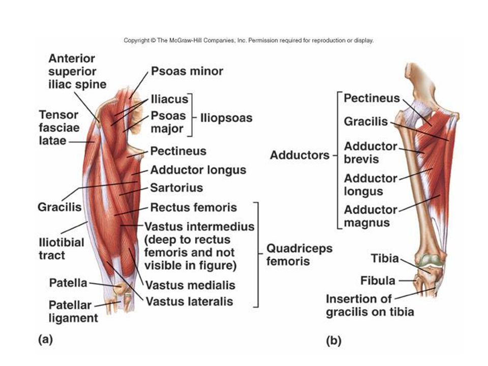
Μύες του Ισχίου Προσαγωγή
Μακρύς / βραχύς / μείζων / ισχνός προσαγωγός, κτενίτης Δυναμική λειτουργία: προσαγωγή κάτω άκρων. ισχυροί εκτείνοντες ή καμπτήρες των ισχίων, ανάλογα με το αν κατά τις κινήσεις κάμψης και έκτασης βρίσκονται μπροστά ή πίσω από τον άξονα περιστροφής (σημαντικός ο ρόλος τους στη βάδιση). Στατική λειτουργία: εξισορρόπηση του ευρισκόμενου σε αστάθεια βάρους του κορμού μέσω της διαρκούς ρύθμισης της θέσης της πυέλου Ενεργοί στην αιώρηση κατά τη βάδιση σκέλος κάτω από Κ.Β. Οι υπόλοιποι συνεισφέρουν σε κάμψη, έξω στροφή ισχίου, κυρίως όταν μηριαίο είναι σε έσω στροφή
. Στατική λειτουργία: εξισορρόπηση του ευρισκόμενου σε αστάθεια βάρους του κορμού μέσω της διαρκούς ρύθμισης της θέσης της πυέλου. Ενεργοί στην αιώρηση κατά τη βάδιση σκέλος κάτω από Κ.Β. Οι υπόλοιποι συνεισφέρουν σε κάμψη, έξω στροφή ισχίου, κυρίως όταν μηριαίο είναι σε έσω στροφή.")
31
Μύες του Ισχίου Προσαγωγή
Κτενίτης Έκφυση: Άνω κλάδος ηβικού οστού. Κατάφυση: Κτενιαία γραμμή του μηριαίου. Νεύρωση: Μηριαίο και θυροειδές νεύρο. Ενέργεια: Προσαγωγή, κάμψη και έξω στροφή Μακρός προσαγωγός Έκφυση: Κάτω από το ηβικό φύμα. Κατάφυση: Μέσο τριτημόριο του έσω σκέλους της τραχείας γραμμής του μηριαίου οστό Νεύρωση: Θυροειδές νεύρο. Ενέργεια: Προσαγωγή και κάμψη ισχίου. Ισχνός προσαγωγός Έκφυση: Χείλος του κάτω κλάδου του ηβικού οστού. Κατάφυση: Κνημιαίο κύρτωμα με τον χήνειο πόδα. Νεύρωση: Θυροειδές νεύρο. Ενέργεια: διαρθρικός μυς, προσαγωγή ισχίου, κάμψη και έσω στροφή γόνατος

32
Μύες του Ισχίου Προσαγωγή
Βραχύς προσαγωγός Έκφυση: Κάτω κλάδος του ηβικού οστού. Κατάφυση: Άνω τριτημόριο τραχείας γραμμής Νεύρωση: Θυροειδές νεύρο. Ενέργεια: Προσαγωγή και έξω στροφή Μέγας προσαγωγός Έκφυση: Κλάδος ισχιακού οστού και κάτω χείλος ισχιακού κυρτώματος. Κατάφυση: Έσω χείλος τραχείας γραμμής και έσω υπερκονδύλιο κύρτωμα μηριαίου οστού. Νεύρωση: Θυροειδές και κνημιαίο νεύρο. Ενέργεια: ισχυρότερος προσαγωγός.

33
Απαγωγοί Μέσος γλουτιαίος Ελάσσων γλουτιαίος Έσω θυροειδής Άνω δίδυμος
Κάτω δίδυμος

34
Μύες του Ισχίου Απαγωγή
Μέσος γλουτιαίος Έκφυση: ΄Εξω χείλος λαγόνιας ακρολοφίας και έξω επιφάνειας λαγόνιου οστού. Κατάφυση: Μείζων τροχαντήρας. Νεύρωση: Άνω γλουτιαίο νεύρο. Ενέργεια: απαγωγή μηρού. Όταν ο μηρός είναι σταθερός (πόδι στήριξης) ενεργώντας ετερόπλευρα κάμπτει τον κορμό πλάγια. Στη βάδιση και στο τρέξιμο ο μυς εμποδίζει από την πλευρά του ποδιού στήριξης την πτώση του άνω μέρους του σώματος προς την πλευρά του κινούμενου ποδιού, διατηρώντας έτσι την κατακόρυφη στάση του κορμού.
 ενεργώντας ετερόπλευρα κάμπτει τον κορμό πλάγια. Στη βάδιση και στο τρέξιμο ο μυς εμποδίζει από την πλευρά του ποδιού στήριξης την πτώση του άνω μέρους του σώματος προς την πλευρά του κινούμενου ποδιού, διατηρώντας έτσι την κατακόρυφη στάση του κορμού.")
35
Μύες του Ισχίου Απαγωγή
Μέσος γλουτιαίος Trendelenburg’s test Η παράλυση του μυός προκαλεί το χήνειο βάδισμα. Απαραίτητη συνεισφορά στην αιώρηση του άλλου σκέλους board.crossfit.com/showthread.php?p=536507 stanford.wellsphere.com/.../manual-muscle-test

36
Μύες του Ισχίου Απαγωγή
Μικρός γλουτιαίος Έκφυση: έξω χείλος λαγόνιας ακρολοφίας. Κατάφυση: Μείζων τροχαντήρας. Νεύρωση: Άνω γλουτιαίο νεύρο. Ενέργεια: Απαγωγή

37
Μύες του Ισχίου Έσω και έξω στροφή μηριαίου
Μύες του Ισχίου Έσω και έξω στροφή μηριαίου Έξω: απιοειδής (1), άνω (2) και κάτω δίδυμος (4), έσω (3) και έξω θυροειδής (5), τετράγωνος μηριαίος (6) Σε κάθε βήμα έχουμε έξω στροφή μηριαίου για να διευκολύνεται η στροφή της λεκάνης Έσω: ελάσσων γλουτιαίος, ΤΠΠ, ημιτενοντώδης, ημιϋμενώδης, μέσος γλουτιαίος Δεν παρουσιάζεται αντίσταση συνήθως έσω στροφείς αδύναμοι (1/3 των έξω)
, άνω (2) και κάτω δίδυμος (4), έσω (3) και έξω θυροειδής (5), τετράγωνος μηριαίος (6) Σε κάθε βήμα έχουμε έξω στροφή μηριαίου για να διευκολύνεται η στροφή της λεκάνης. Έσω: ελάσσων γλουτιαίος, ΤΠΠ, ημιτενοντώδης, ημιϋμενώδης, μέσος γλουτιαίος. Δεν παρουσιάζεται αντίσταση συνήθως έσω στροφείς αδύναμοι (1/3 των έξω)")
38
Απιοειδής Έκφυση: πρόσθια επιφάνεια ιερού οστού
Κατάφυση: μείζων τροχαντήρας Νεύρωση: Ι1-2 Ενέργεια: Έξω στροφή σε έκταση, έσω στροφή σε κάμψη, έκταση, απαγωγή

39
Μύες του Ισχίου Οριζόντια απαγωγή και προσαγωγή
Μύες του Ισχίου Οριζόντια απαγωγή και προσαγωγή Συμβαίνει όταν το ισχίο είναι σε κάμψη 90°, με μηριαίο σε απαγωγή ή προσαγωγή Απαιτείται συντονισμένη δράση Οι οπίσθιοι μύες αποτελεσματικότεροι ως οριζόντιοι απαγωγοί και προσαγωγοί από τους πρόσθιους Οι οπίσθιοι διατείνονται όταν το μηριαίο είναι σε κάμψη 90°, ενώ στους πρόσθιους η τάση μειώνεται

41
Νεύρα Ισχιακό νεύρο Το μεγαλύτερο στον άνθρωπο
Συνεχίζει σε κνημιαίο και περονιαίο νεύρο. Σχηματίζεται από πρόσθιους κλάδους Ο4-Ι3 νεύρων και είναι συνέχεια ιερού πλέγματος Νευρώνει την άρθρωση του ισχίου, το δικέφαλο μηριαίο, ημιτενοντώδη, ημιυμενώδη και ισχιακή κεφαλή μείζονα προσαγωγού Sciatic Nerve The sciatic nerve (image) is the largest nerve in the body, and consists of the medially placed tibial nerve and the laterally placed common peroneal nerve. It is formed from the ventral rami of the fourth lumbar to third sacral spinal nerves and is a continuation of the upper band of the sacral plexus. It leaves the pelvis through the greater sciatic foramen, below the piriformis muscle (animation) , and descends between the greater trochanter of the femur and the ischial tuberosity. Initially deep to piriformis, it runs inferiorly and laterally posterior to the ischium, crossing over the nerve to quadratus femoris. Inferior to piriformis; it lies deep to gluteus maximus. It passes inferiorly crossing obturator internus, the gemelli and quadratus femoris. The posterior cutaneous nerve of thigh (image) and the inferior gluteal artery lie on its medial side. Descending vertically, it enters the thigh at the lower border of gluteus maximus, where it lies on the posterior surface of adductor magnus (animation) . It gives off nerves to the hamstring muscles. The nerve is crossed obliquely on its superficial aspect by the long head of biceps femoris (image) . The nerve ends at the upper aspect of the popliteal fossa by dividing into the tibial and common perineal nerves. The nerve can be represented on the back of the thigh by a line drawn from just medial to the midpoint of the line from the ischial tuberosity to the apex of greater trochanter down to the apex of popliteal fossa. It supplies articular branches to the hip joint, with muscular branches to biceps femoris, semitendinosus and semimembranosus and the ischial head of adductor magnus. The nerve to the short head of biceps is from the common peroneal division, with the other muscular branches emerging from the tibial division. 41
 is the largest nerve in the body, and consists of the medially placed tibial nerve and the laterally placed common peroneal nerve. It is formed from the ventral rami of the fourth lumbar to third sacral spinal nerves and is a continuation of the upper band of the sacral plexus. It leaves the pelvis through the greater sciatic foramen, below the piriformis muscle (animation) , and descends between the greater trochanter of the femur and the ischial tuberosity. Initially deep to piriformis, it runs inferiorly and laterally posterior to the ischium, crossing over the nerve to quadratus femoris. Inferior to piriformis; it lies deep to gluteus maximus. It passes inferiorly crossing obturator internus, the gemelli and quadratus femoris. The posterior cutaneous nerve of thigh (image) and the inferior gluteal artery lie on its medial side. Descending vertically, it enters the thigh at the lower border of gluteus maximus, where it lies on the posterior surface of adductor magnus (animation) . It gives off nerves to the hamstring muscles. The nerve is crossed obliquely on its superficial aspect by the long head of biceps femoris (image) . The nerve ends at the upper aspect of the popliteal fossa by dividing into the tibial and common perineal nerves. The nerve can be represented on the back of the thigh by a line drawn from just medial to the midpoint of the line from the ischial tuberosity to the apex of greater trochanter down to the apex of popliteal fossa. It supplies articular branches to the hip joint, with muscular branches to biceps femoris, semitendinosus and semimembranosus and the ischial head of adductor magnus. The nerve to the short head of biceps is from the common peroneal division, with the other muscular branches emerging from the tibial division. 41.")
42
Νεύρα Μηριαίο νεύρο λαγονοσφυικό πλέγμα (Ο2,3,4) τετρακέφαλος, λαγονοψοΐτης, κτενίτης. Νευρώνει όλους τους μυς της πρόσθιας επιφάνειας μηρού Θυροειδές νεύρο βραχύς προσαγωγός, μακρός, ισχνός προσαγωγός + αισθητικό κλάδο Femoral Nerve Anatomy Text The femoral nerve (image) is a branch of the lumbosacral plexus. It arises from the posterior divisions of the ventral rami of the second, third and fourth lumbar nerves. It passes infero-laterally through the substance of psoas major (animation) , behind the obturator nerve, gaining the groove between the psoas major and iliacus muscles, just below the iliac crest. It descends in the groove behind the iliac fascia. Within the abdomen, it gives off branches to iliacus, pectineus and the femoral artery. The nerve (or nerves) to pectineus arises close to the inguinal ligament, passing behind the femoral vessels to the lateral border of the muscle. The nerve enters the thigh posterior to the inguinal ligament and lateral to the femoral artery and femoral sheath. It divides into anterior and posterior divisions. It supplies all the muscles in the anterior compartment of the thigh (image) . It also gives articular branches to the hip and knee joints. A branch to the hip joint arises from the nerve to rectus femoris and branches to the knee joint arise from each of the nerves supplying the vastus muscles with a fourth branch possibly arising from the saphenous nerve Obturator Nerve The obturator nerve (image) arises from the ventral divisions of the ventral rami of the second, third and fourth lumbar nerves. It descends through psoas major, emerging from its medial border at the pelvic brim. It passes behind the common iliac vessels and descends lateral to the internal iliac vessels, along the lateral wall of the true pelvis, where it lies on the obturator internus muscle (image) . It enters the thigh through the upper part of the obturator foramen. Here it separates into anterior and posterior branches. The anterior branch descends (image) in front of obturator externus and adductor brevis and behind pectineus and adductor longus. It gives a branch to the hip joint as it enters the thigh. Branches to adductor longus, gracilis, adductor brevis and sometimes pectineus, arise as the nerve descends between the muscle layers. At the lower border of adductor longus, it contributes a branch to the subsartorial plexus. Terminally, it gives off a vascular and sometimes a cutaneous branch. When present, the cutaneous branch passes between gracilis and adductor longus to supply the skin over the lower two-thirds of the medial side of the thigh. The posterior branch penetrates and supplies the obturator externus muscle, and then descends between adductor brevis and magnus. It penetrates adductor magnus, supplying its upper part, to enter the popliteal fossa behind the popliteal artery. It supplies the posterior aspect of the knee joint and the cruciate ligaments. 42
 is a branch of the lumbosacral plexus. It arises from the posterior divisions of the ventral rami of the second, third and fourth lumbar nerves. It passes infero-laterally through the substance of psoas major (animation) , behind the obturator nerve, gaining the groove between the psoas major and iliacus muscles, just below the iliac crest. It descends in the groove behind the iliac fascia. Within the abdomen, it gives off branches to iliacus, pectineus and the femoral artery. The nerve (or nerves) to pectineus arises close to the inguinal ligament, passing behind the femoral vessels to the lateral border of the muscle. The nerve enters the thigh posterior to the inguinal ligament and lateral to the femoral artery and femoral sheath. It divides into anterior and posterior divisions. It supplies all the muscles in the anterior compartment of the thigh (image) . It also gives articular branches to the hip and knee joints. A branch to the hip joint arises from the nerve to rectus femoris and branches to the knee joint arise from each of the nerves supplying the vastus muscles with a fourth branch possibly arising from the saphenous nerve. Obturator Nerve. The obturator nerve (image) arises from the ventral divisions of the ventral rami of the second, third and fourth lumbar nerves. It descends through psoas major, emerging from its medial border at the pelvic brim. It passes behind the common iliac vessels and descends lateral to the internal iliac vessels, along the lateral wall of the true pelvis, where it lies on the obturator internus muscle (image) . It enters the thigh through the upper part of the obturator foramen. Here it separates into anterior and posterior branches. The anterior branch descends (image) in front of obturator externus and adductor brevis and behind pectineus and adductor longus. It gives a branch to the hip joint as it enters the thigh. Branches to adductor longus, gracilis, adductor brevis and sometimes pectineus, arise as the nerve descends between the muscle layers. At the lower border of adductor longus, it contributes a branch to the subsartorial plexus. Terminally, it gives off a vascular and sometimes a cutaneous branch. When present, the cutaneous branch passes between gracilis and adductor longus to supply the skin over the lower two-thirds of the medial side of the thigh. The posterior branch penetrates and supplies the obturator externus muscle, and then descends between adductor brevis and magnus. It penetrates adductor magnus, supplying its upper part, to enter the popliteal fossa behind the popliteal artery. It supplies the posterior aspect of the knee joint and the cruciate ligaments. 42.")
43
Βιομηχανική Κάμψη: 110° - 120 ° Eκταση: 10 ° -15 ° Aπαγωγή: 30 °- 50 °
Προσαγωγή: 30 ° Έσω Στροφή:40 ° - 60 ° Έξω Στροφή: 30 °- 40 ° Magee (1997) Κλειδωμένη /σφικτή θέση άρθρωσης: πλήρης έκταση, έσω στροφή & απαγωγή Χαλαρή θέση άρθρωσης: 30°κάμψη, 30° απαγωγή & μερική έξω στροφή. Πότε χρειάζεται; Σε φλεγμονή -μειώνει τον πόνο και επιτρέπει τη δημιουργία οιδήματος
 Κλειδωμένη /σφικτή θέση άρθρωσης: πλήρης έκταση, έσω στροφή & απαγωγή. Χαλαρή θέση άρθρωσης: 30°κάμψη, 30° απαγωγή & μερική έξω στροφή. Πότε χρειάζεται; Σε φλεγμονή -μειώνει τον πόνο και επιτρέπει τη δημιουργία οιδήματος.")
44
Φορτίσεις στην άρθρωση του ισχίου
Το ισχίο παίζει σημαντικό ρόλο στη στήριξη του Σ.Β. Η γραμμή του ΚΒ βρίσκεται πίσω από ηβική σύμφυση Όταν το βάρος είναι όμοια κατανεμημένο στα δύο πόδια κατά την όρθια στάση, το βάρος που υποδέχεται κάθε ισχίο είναι ίσο με το 1/2 του βάρους των μελών του σώματος πάνω από το ισχίο, ή περίπου ίσο με το 1/3 του συνολικού Σ.Β.. Η συνολική επιβάρυνση σε κάθε ισχίο θα είναι μεγαλύτερη από την προαναφερόμενη, επειδή η τάση στους μεγάλους μυς του ισχίου προσθέτει επιπλέον συμπιεστική δύναμη στην άρθρωση (κυρίως μετά από πολύωρη ορθοστασία)
")
45
Φορτίσεις στην άρθρωση του ισχίου
Οι συμπιεστικές δυνάμεις στο ισχίο στη φάση στήριξης στο βάδισμα είναι 2,38 φορές το σωματικού βάρους (ΣΒ), κατά την ανάβαση σκαλοπατιών 2,51 ΣΒ, στο αργό τρέξιμο 5,20 ΣΒ Στη μονοποδική στήριξη το ΚΒ μετατοπίζεται σε όλα τα επίπεδα. Αυξάνονται οι δυνάμεις και παραγόμενες ροπές ανάλογα με στάση σώματος, θέση αιωρούμενου ποδιού και άνω άκρων και κλίση λεκάνης Στην αιώρηση η συμπίεση είναι ίση με Σ.Β. Η επιβάρυνση είναι αυξημένη στη στήριξη κατά τη διάρκεια αθλητικών δραστηριοτήτων, όταν φοράμε σκληρά υποδήματα, κατά τη μεταφορά βάρους, όταν αυξάνει η ταχύτητα βάδισης ή τρεξίματος, κλπ
, κατά την ανάβαση σκαλοπατιών 2,51 ΣΒ, στο αργό τρέξιμο 5,20 ΣΒ. Στη μονοποδική στήριξη το ΚΒ μετατοπίζεται σε όλα τα επίπεδα. Αυξάνονται οι δυνάμεις και παραγόμενες ροπές ανάλογα με στάση σώματος, θέση αιωρούμενου ποδιού και άνω άκρων και κλίση λεκάνης. Στην αιώρηση η συμπίεση είναι ίση με Σ.Β. Η επιβάρυνση είναι αυξημένη στη στήριξη κατά τη διάρκεια αθλητικών δραστηριοτήτων, όταν φοράμε σκληρά υποδήματα, κατά τη μεταφορά βάρους, όταν αυξάνει η ταχύτητα βάδισης ή τρεξίματος, κλπ.")
46
Πρέπει να γνωρίζετε: Όλους τους μυς (όχι μόνο αυτούς που καλύψαμε σήμερα) Έκφυση- κατάφυση, νεύρωση Οστικές προεξοχές- και τι προσφύεται εκεί Νεύρα- τι νευρώνουν Σύνδεσμοι: έκφυση- κατάφυση, λειτουργία

47
Βιβλιογραφία Hall Susan J. (2005). Εμβιομηχανική. Εκδόσεις Παρισιάνου, Αθήνα. Weineck Jurgen (1998). Ανατομική της άθλησης. Εκδόσεις Σάλτο, Θεσσαλονίκη. Hamilton N., Luttgens K. (2003). Κινησιολογία. Εκδόσεις Παρισιάνου, Αθήνα. Robertson G., Caldwell G., Hamill J., Kamen G., Whittlesey S. (2004). Research Methods in Biomechanics. Human Kinetics, Champaign, IL. Anatomy t.v. Ppt
. Ανατομική της άθλησης. Εκδόσεις Σάλτο, Θεσσαλονίκη. Hamilton N., Luttgens K. (2003). Κινησιολογία. Εκδόσεις Παρισιάνου, Αθήνα. Robertson G., Caldwell G., Hamill J., Kamen G., Whittlesey S. (2004). Research Methods in Biomechanics. Human Kinetics, Champaign, IL. Anatomy t.v. Ppt.")
Παρόμοιες παρουσιάσεις