Κατέβασμα παρουσίασης
Η παρουσίαση φορτώνεται. Παρακαλείστε να περιμένετε

1
Ανατομία dsfsf dsfsf Ισχίο dsfsf Ελευθερία Θωμαΐδου, Pt

2
Μαθησιακά αποτελέσματα
Στο τέλος του μαθήματος πρέπει να είστε σε θέση να : Αναγνωρίζετε τα οστά που σχηματίζουν την άρθρωση του ισχίου και να εντοπίζετε τα οστικά σημεία Κατανοείτε τις κινήσεις της άρθρωσης του ισχίου Εντοπίζετε τους συνδέσμους, θύλακα,νεύρα Αναγνωρίζετε τις προσφύσεις των μυών που δρουν στην περιοχή

3
Άρθρωση Ισχίου-Hip joint
Η κοτύλη υποδέχεται την κεφαλή του μηριαίου

4
Άρθρωση Ισχίου

5
Μηριαίο οστό -Femur Είναι το μακρύτερο οστό του σώματος ( 34-53 εκατ)
Διακρίνουμε 2 επιφύσεις και 1 διάφυση( shaft) Γλουτιαίο τράχυσμα Gluteal tuberosity Τραχεία γραμμή Linea aspera Έσω υπερκονδύλια γραμμή Med. supracondylar line Έξω υπερκονδύλια γραμμή Lat. supracondylar line Ιγνυακή επιφάνεια Popliteal surface
 Γλουτιαίο τράχυσμα Gluteal tuberosity. Τραχεία γραμμή Linea aspera. Έσω υπερκονδύλια γραμμή Med. supracondylar line. Έξω υπερκονδύλια γραμμή Lat. supracondylar line. Ιγνυακή επιφάνεια Popliteal surface.")
6
Η κεφαλή αποτελεί τα 2/3 σφαίρας, στο κέντρο της οποίας υπάρχει κοίλωμα, ο βόθρος της κεφαλής όπου προσφύεται ο στρογγύλος σύνδεσμος

7
Μηριαίο Οστό Femur Μηριαία Κεφαλή Femoral head
Αυχένας Neck -χειρουργικός - ανατομικός Μείζων τροχαντήρας Greater trochanter Έλασσων τροχαντήρας Lesser trochanter FH GT N LT

8
ο αυχένας είναι οστέινος κύλινδρος
Υπό γωνία 125ο Ο προσανατολισμός του αυχένα σε σχέση με τη διάφυση αυξάνει το εύρος των κινήσεων

9
Ο μείζων τροχαντήρας φέρει το τροχαντήριο βόθρο
Και ο έλασσων τροχαντήρας βρίσκεται πιο κάτω και έχει αμβλύ κωνικό σχήμα Από τα χείλη του μ.τροχαντήρα φέρονται η πρόσθια και οπίσθια μεσοτροχαντήρια γραμμή Το μηριαίο παρουσιάζει 3 επιφάνειες ( πρόσθια, έσω και έξω) και τρία χείλη ( έσω ,έξω και οπίσθιο)
 και τρία χείλη ( έσω ,έξω και οπίσθιο)")
10
Το οπίσθιο χείλος ονομάζεται τραχεία γραμμή
Η τραχεία γραμμή έχει δύο χείλη Γλουτιαίο τράχυσμα-> πρόσφυση μεγ.γλουτιαίου Κτενιαία γραμμή-> κατάφυση κτενίτη μυ

11
Οστική Ψηλάφηση ASIS Iliac crest PSIS Ischial tuberosity Sacrum
Greater trochanter Pubic bones (pubic tubercles)
")
12
Hip Extension Hip Flexion

13
Hip Medial Rotation Hip Lateral Rotation

14
Hip Adduction opposite
Hip Abduction shown Adduction

15
Joints of the Pelvic Girdle
Hip (coxal) Joint Sacroiliac Joint (SIJ) Pubic Symphysis
 Joint. Sacroiliac Joint (SIJ) Pubic Symphysis.")
16
Hip (coxal) Joint Διάρθρωση synovial joint
Μηνοειδής επιφάνεια της κοτύλης συμπληρώνεται από τον κοτυλιαίο δακτύλιο οπ οποίος σχηματίζει πάνω από την κοτυλιαία εντομή, τον εγκάρσιο σύνδεσμο και κεφαλή μηριαίου Spherical femoral head Βαθειά κοτύλη Deep acetabulum Σταθερότητα Περιορισμένο ROM

17
The Sacroiliac Joint Synovial planar joint Posterior joint
Limited movement

18
Σύνδεσμοι Ισχίου Ο λαγονιομηρικός-Iliofemoral Ligament
(ΠΚΛΑ- πρόσθια μεσοτροχαντήρια γραμμή) Ο ηβομηρικός –pubofemoral ligament (θυροειδής ακρολοφία- πρόσθια μεσοτροχαντήρια γραμμή) Ο ισχιομηρικός –ischiofemoral ligament ( όφρυ της κοτύλης- τροχαντήριος βόθρος) Ο στρογγύλος( μηνοειδής επιφάνεια κοτύλης- βόθρος μηριαίας κεφαλής)
 Ο ηβομηρικός –pubofemoral ligament. (θυροειδής ακρολοφία- πρόσθια μεσοτροχαντήρια γραμμή) Ο ισχιομηρικός –ischiofemoral ligament. ( όφρυ της κοτύλης- τροχαντήριος βόθρος) Ο στρογγύλος( μηνοειδής επιφάνεια κοτύλης- βόθρος μηριαίας κεφαλής)")
19
Ligaments of the Hip and Pelvis
Inguinal Ligament Iliofemoral Ligament Pubofemoral Ligament

20
Ligaments of the Hip and Pelvis
Ischiofemoral Ligament

21
Βουβωνικός σύνδεσμος Βρίσκεται μπροστά από το λαγόνιο, ψοϊτη, κτενίτη, αγγεία και νεύρα. Δίνει προσφύσεις στην περιτονία, στον έσω λοξό και στο ν εγκάρσιο The pubofemoral ligament is a triangular band; the base is attached to the iliopubic eminence, superior pubic ramus and the pubic part of the acetabular rim. It passes distally to blend with the inferior aspect of the hip joint capsule and underside of the femoral neck. Like the iliofemoral ligament, it is taut in extension. The iliofemoral ligament is a broad thick triangular band. It is attached proximally to the anterior inferior iliac spine and acetabular rim and distally to the intertrochanteric line. The ligament is thicker towards the edges and thinner in the middle. Its medial band is vertically orientated, and its lower end attaches to the medial end of the intertrochanteric line. The lateral band is obliquely orientated and is attached to a tubercle at the upper lateral part of the intertrochanteric line. This is often considered as an inverted Y- or V-shaped structure; the lateral band may then be referred to as the ‘iliotrochanteric ligament’. It is taut in extension. The inguinal ligament lies anterior to the structures passing from the abdomen (image) into the lower limb, these include the iliacus, psoas and pectineus muscles, the femoral canal, vessels and nerve. The ligament gives attachment along its whole length to the transversalis fascia. The iliac fascia, internal oblique and transversus abdominis muscles are attached to its lateral part.
 into the lower limb, these include the iliacus, psoas and pectineus muscles, the femoral canal, vessels and nerve. The ligament gives attachment along its whole length to the transversalis fascia. The iliac fascia, internal oblique and transversus abdominis muscles are attached to its lateral part.")
22
Λαγονομηρικός •Ο ισχυρότερος του ανθρώπινου σώματος
•Από τη ΠΚΛΑ και το χείλος της κοτύλης στη μεσοτροχαντήρια γραμμή •Έχει σχήμα V •Είναι πιο παχύς στα άκρα και πιο λεπτός στο μέσο. •Περιορίζει έκταση και έξω στροφή •Το κατακόρυφο τμήμα του κατά την όρθια στάση του σώματος εμποδίζει την πτώση του κορμού προς τα πίσω, ενώ το οριζόντιο τμήμα κατά τη βάδιση εμποδίζει τη βύθιση του άνω μέρους του σώματος προς την πλευρά του ελεύθερου κάτω άκρου

23
Ηβομηρικός •Είναι μια τριγωνική δέσμη
•Η βάση του είναι συνδεδεμένη με τον άνω κλάδο ηβικού οστού και το ηβικό τμήμα της κοτύλης. •Συνδέεται με την κάτω πλευρά του αρθρικού θύλακα και με την κάτω πλευρά του αυχένα του μηριαίου •Περιορίζει την έκταση και απαγωγή

24
Ισχιομηρικός σύνδεσμος
Από το ισχιακό τμήμα της κοτύλης και πίσω από την άρθρωση του ισχίου •Συγκλίνουν στην άνω έξω πλευρά του αυχένα του μηριαίου •Περιορίζει έκταση και έσω στροφή Ι

25
Ισχιοιερός σύνδεσμος ΟΑΛΑ, ΟΚΛΑ, οπίσθια επιφάνεια ιερού (όπου μπλέκεται με ιερολαγόνιο σύνδεσμο), κόκκυγας •Οι ίνες συγκλίνουν καθώς διέρχονται κάτω και πλάγια. Στρέφονται και μετά αποκλίνουν για να συνδεθούν στο ισχιακό κύρτωμα και τον κάτω ισχιακό κλάδο •Εμποδίζει οπίσθια στροφή λαγονίου •Αποτελεί πηγή πόνου The ischiofemoral ligament (image) arises from the ischial component of the acetabulum below and behind the hip joint. The fibers pass horizontally behind the joint, converging on the upper lateral aspect of the femoral neck. The sacrotuberous ligament (image) has an extensive attachment; to the posterior superior and posterior inferior iliac spines, the posterior surface of the sacrum (where it blends with the posterior (dorsal) sacro-iliac ligaments), the lateral aspect of the lower sacrum and to the upper surface of the coccyx. The fibers converge as they pass downwards and laterally. They twist on themselves and then diverge to attach to the medial margin of the ischial tuberosity and the lower margin of the ischial ramus. Distal fibers of gluteus maximus attach to the ligaments posterior surface.
, κόκκυγας •Οι ίνες συγκλίνουν καθώς διέρχονται κάτω και πλάγια. Στρέφονται και μετά αποκλίνουν για να συνδεθούν στο ισχιακό κύρτωμα και τον κάτω ισχιακό κλάδο •Εμποδίζει οπίσθια στροφή λαγονίου. •Αποτελεί πηγή πόνου. The ischiofemoral ligament (image) arises from the ischial component of the acetabulum below and behind the hip joint. The fibers pass horizontally behind the joint, converging on the upper lateral aspect of the femoral neck. The sacrotuberous ligament (image) has an extensive attachment; to the posterior superior and posterior inferior iliac spines, the posterior surface of the sacrum (where it blends with the posterior (dorsal) sacro-iliac ligaments), the lateral aspect of the lower sacrum and to the upper surface of the coccyx. The fibers converge as they pass downwards and laterally. They twist on themselves and then diverge to attach to the medial margin of the ischial tuberosity and the lower margin of the ischial ramus. Distal fibers of gluteus maximus attach to the ligaments posterior surface.")
26
Στρογγύλος σύνδεσμος/σύνδεσμος της κεφαλής του μηριαίου
Τριγωνική, επίπεδη δεσμίδα •Είναι σφιχτός όταν ο μηρός είναι σχεδόν σε κάμψη και προσαγωγή •Ποικίλει σε πάχος και ισχύ •Σε συνέχεια με μια αρτηρία για την παροχή αίματος στη κεφαλή του μηριαίου he ligamentum teres, or ligament of the head of the femur, is a triangular flattened band. The apex attaches to the pit or fovea on the head of the femur and two bands attach the base onto either side of the acetabular notch. Between the bony attachments the ligament blends with the transverse acetabular ligament. It lies in a synovial sheath. The ligament is taut when the thigh is semi-flexed and adducted. The ligament varies in size and rarely may be absent. A small artery is present within its substance. After closure of the growth plate, there is an anastomosis between this artery and the metaphyseal vessels.

27
Ο επιχείλιος χόνδρος Λειτουργίες Αυξάνει το βάθος της κοτύλης
Βελτιώνει την αρνητική ενδο-αρθρική πίεση Παρέχει σταθερότητα Articular Hyaline Cartilage Anatomy Text Jump to Clinical Pathology Text The articular surfaces of the head of the femur, the medial and lateral femoral condyles, the lunate surface, pubic symphysis and the grooved surface of the lesser sciatic notch of the hip bone, the auricular surfaces of the hip bone and sacrum, the tibial plateau of the tibia and the posterior aspect of the patella, are covered by a variety of hyaline cartilage termed ‘articular hyaline cartilage’. Its protein content distinguishes articular hyaline cartilage; it contains Type II collagen only. It offers a firm, smooth and relatively friction-free surface facilitating joint movements. Articular hyaline cartilage possesses a degree of compressibility and elasticity. These features enable the articular surfaces to dissipate laterally the vertical compressive forces to which the joints are subjected during weight transmission. Articular hyaline cartilage does not usually ossify. Intra-Articular Fat Pad Anatomy Text The acetabular fossa is a shallow depression in the floor of the acetabulum, above the acetabular notch. The fossa is lined with a fibroelastic fat pad, which in turn is covered with synovial membrane. The membrane is attached to the medial aspect of the transverse ligament and the margins of the acetabular fossa, enveloping the ligament of the head of the femur, where it extends up to the edge of the pit (fovea) on the femoral head. Ligamentum Teres (Syn. Femoral Head Ligament) Anatomy Text Jump to Clinical Pathology Text The ligamentum teres (image) , or ligament of the head of the femur, is a triangular flattened band. The apex attaches to the pit or fovea (image) on the head of the femur and two bands attach the base onto either side of the acetabular notch. Between the bony attachments the ligament blends with the transverse acetabular ligament (image) . It lies in a synovial sheath. The ligament is taut when the thigh is semi-flexed and adducted. Clinical Pathology Text Jump to Anatomy Text The ligamentum teres originates from the acetabular notch. Its exact function is unclear but increasing experience from hip arthroscopy suggests that it may be abnormal in certain pathological states. The ligament varies in size and rarely may be absent. A small artery is present within its substance. After closure of the growth plate, there is an anastomosis between this artery and the metaphyseal vessels. Arteries The blood supply of the hip changes with growth. The main blood supply in the adult originates from the medial and lateral femoral circumflex arteries. The obturator artery and inferior and superior circumflex arteries also demonstrate some blood supply to the hip joint. There is a vessel within the ligamentum teres known as the artery of the ligamentum teres. This may contribute some blood supply to the epiphyseal region of the femoral head but its significance is otherwise unknown. The femoral head is supplied by three terminal arterial sources: the artery of the ligamentum teres, a terminal branch of the lateral femoral circumflex artery and the terminal branch of the medial circumflex artery, the lateral epiphyseal artery. The latter is the critical blood supply to the majority of the weight bearing superior portion of the femoral head. This artery is particularly at risk during posterior fracture dislocation. The adult femoral head ranges in diameter from 40mm to 60mm and is not a perfect sphere. Its subtle ace veracity is reflected on the acetabular side. Accurate reduction of femoral head fragments is necessary in order to maximize contact area between the femoral head and the acetabulum and to minimize stresses across the articular cartilage. The management of femoral head fractures involves adequate imaging with plain X-rays and CT scans followed by open reduction and internal fixation. The specific procedure undertaken will depend on any associated hip instability and/or acetabular injuries. The blood supply of the femoral head changes with age. Before the age of four, the metaphyseal and lateral epiphyseal blood vessels supply the majority of the blood supply. After the age of four, the only blood supply of note comes from the lateral epiphyseal blood vessels. The adolescent’s blood supply improves again when the metaphyseal blood supply returns. Given the tenuous blood supply to the femoral head in children, the incidence of osteonecrosis after fracture is 20 to 45% compared to 10% in the general population. Acetabular Labrum Anatomy Text Jump to Clinical Pathology Text The hip joint is an extraordinarily stable joint. Its stability is related primarily to the bony and labral anatomy of the acetabulum and femoral head. This is greatly supplemented by the thick fibrous capsule of the hip with its ligamentous condensations and local muscular anatomy. A fibrocartilaginous rim, the acetabular labrum (image) , increases the depth of the labrum. This is firmly attached to the bony acetabular rim and to the transverse acetabular ligament. The free edge of the labrum forms a smaller circle than the base, clasping the head of the femur (image) . The femoral head forms approximately two-thirds of a sphere and is situated on the femoral neck approximately three-quarters of the diameter of the femoral head. This relationship in size between the femoral head and femoral neck allows the femoral head to be deeply seated within its acetabular socket without compromising the stability or range of motion. The acetabular labrum (animation) serves to deepen the acetabulum and increase the stability of the joint. The addition of the labrum ensures that at least 50% of the femoral head is covered by osteocartilaginous labral acetabular complex in any position of hip motion. A normal hip (movie) will typically extend 20 to 30 degrees, flex 135 degrees, abduct 45 to 50 degrees, adduct 20 to 30 degrees and have a combined arc rotation of 90 degrees. The extent of internal external rotation may depend on the amount of anteversion of the femoral neck. Moreover the rotation of the hip may differ if it is tested in flexion or extension. This is particularly the case if there is an abnormality of femoral head shape. Transverse Acetabular Ligament Anatomy Text Jump to Clinical Pathology Text The transverse acetabular ligament (image) bridges the acetabular notch, converting it into a foramen. The superficial edge of the ligament is at the level of the acetabular rim, forming part of the labrum. The foramen deep to the ligament transmits articular vessels and nerves. Clinical Pathology Text Pubofemoral Ligament Anatomy Text A capsule that extends posteriorly from the acetabulum to the mid-femoral neck and anteriorly from the acetabulum to the intra-trochanteric ridge surrounds the hip joint. There are a number of condensations of capsular fibers known as: 3) The ischiofemoral Ligament. 2) The pubofemoral Ligament. 1) The iliofemoral Ligament. The pubofemoral ligament is a triangular band; the base is attached to the iliopubic eminence, superior pubic ramus and the pubic part of the acetabular rim. It passes distally to blend with the inferior aspect of the hip joint capsule and underside of the femoral neck. Like the iliofemoral ligament, it is taut in extension. Ischiofemoral Ligament Anatomy Text A capsule that extends posteriorly from the acetabulum to the mid-femoral neck and anteriorly from the acetabulum to the intra-trochanteric ridge surrounds the hip joint. There are a number of condensations of capsular fibers known as: The ischiofemoral ligament (image) arises from the ischial component of the acetabulum below and behind the hip joint. The fibers pass horizontally behind the joint, converging on the upper lateral aspect of the femoral neck. Iliofemoral Ligament Anatomy Text Jump to Clinical Pathology Text A capsule that extends posteriorly from the acetabulum to the mid-femoral neck and anteriorly from the acetabulum to the intra-trochanteric ridge surrounds the hip joint. There are a number of condensations of capsular fibers known as: The iliofemoral ligament is a broad thick triangular band. It is attached proximally to the anterior inferior iliac spine and acetabular rim and distally to the intertrochanteric line. The ligament is thicker towards the edges and thinner in the middle. Its medial band is vertically orientated, and its lower end attaches to the medial end of the intertrochanteric line. The lateral band is obliquely orientated and is attached to a tubercle at the upper lateral part of the intertrochanteric line. This is often considered as an inverted Y- or V-shaped structure; the lateral band may then be referred to as the ‘iliotrochanteric ligament’. It is taut in extension.
 on the femoral head. Ligamentum Teres (Syn. Femoral Head Ligament) Anatomy Text Jump to Clinical Pathology Text The ligamentum teres (image) , or ligament of the head of the femur, is a triangular flattened band. The apex attaches to the pit or fovea (image) on the head of the femur and two bands attach the base onto either side of the acetabular notch. Between the bony attachments the ligament blends with the transverse acetabular ligament (image) . It lies in a synovial sheath. The ligament is taut when the thigh is semi-flexed and adducted. Clinical Pathology Text Jump to Anatomy Text The ligamentum teres originates from the acetabular notch. Its exact function is unclear but increasing experience from hip arthroscopy suggests that it may be abnormal in certain pathological states. The ligament varies in size and rarely may be absent. A small artery is present within its substance. After closure of the growth plate, there is an anastomosis between this artery and the metaphyseal vessels. Arteries The blood supply of the hip changes with growth. The main blood supply in the adult originates from the medial and lateral femoral circumflex arteries. The obturator artery and inferior and superior circumflex arteries also demonstrate some blood supply to the hip joint. There is a vessel within the ligamentum teres known as the artery of the ligamentum teres. This may contribute some blood supply to the epiphyseal region of the femoral head but its significance is otherwise unknown. The femoral head is supplied by three terminal arterial sources: the artery of the ligamentum teres, a terminal branch of the lateral femoral circumflex artery and the terminal branch of the medial circumflex artery, the lateral epiphyseal artery. The latter is the critical blood supply to the majority of the weight bearing superior portion of the femoral head. This artery is particularly at risk during posterior fracture dislocation. The adult femoral head ranges in diameter from 40mm to 60mm and is not a perfect sphere. Its subtle ace veracity is reflected on the acetabular side. Accurate reduction of femoral head fragments is necessary in order to maximize contact area between the femoral head and the acetabulum and to minimize stresses across the articular cartilage. The management of femoral head fractures involves adequate imaging with plain X-rays and CT scans followed by open reduction and internal fixation. The specific procedure undertaken will depend on any associated hip instability and/or acetabular injuries. The blood supply of the femoral head changes with age. Before the age of four, the metaphyseal and lateral epiphyseal blood vessels supply the majority of the blood supply. After the age of four, the only blood supply of note comes from the lateral epiphyseal blood vessels. The adolescent’s blood supply improves again when the metaphyseal blood supply returns. Given the tenuous blood supply to the femoral head in children, the incidence of osteonecrosis after fracture is 20 to 45% compared to 10% in the general population. Acetabular Labrum Anatomy Text Jump to Clinical Pathology Text The hip joint is an extraordinarily stable joint. Its stability is related primarily to the bony and labral anatomy of the acetabulum and femoral head. This is greatly supplemented by the thick fibrous capsule of the hip with its ligamentous condensations and local muscular anatomy. A fibrocartilaginous rim, the acetabular labrum (image) , increases the depth of the labrum. This is firmly attached to the bony acetabular rim and to the transverse acetabular ligament. The free edge of the labrum forms a smaller circle than the base, clasping the head of the femur (image) . The femoral head forms approximately two-thirds of a sphere and is situated on the femoral neck approximately three-quarters of the diameter of the femoral head. This relationship in size between the femoral head and femoral neck allows the femoral head to be deeply seated within its acetabular socket without compromising the stability or range of motion. The acetabular labrum (animation) serves to deepen the acetabulum and increase the stability of the joint. The addition of the labrum ensures that at least 50% of the femoral head is covered by osteocartilaginous labral acetabular complex in any position of hip motion. A normal hip (movie) will typically extend 20 to 30 degrees, flex 135 degrees, abduct 45 to 50 degrees, adduct 20 to 30 degrees and have a combined arc rotation of 90 degrees. The extent of internal external rotation may depend on the amount of anteversion of the femoral neck. Moreover the rotation of the hip may differ if it is tested in flexion or extension. This is particularly the case if there is an abnormality of femoral head shape. Transverse Acetabular Ligament Anatomy Text Jump to Clinical Pathology Text The transverse acetabular ligament (image) bridges the acetabular notch, converting it into a foramen. The superficial edge of the ligament is at the level of the acetabular rim, forming part of the labrum. The foramen deep to the ligament transmits articular vessels and nerves. Clinical Pathology Text. Pubofemoral Ligament Anatomy Text A capsule that extends posteriorly from the acetabulum to the mid-femoral neck and anteriorly from the acetabulum to the intra-trochanteric ridge surrounds the hip joint. There are a number of condensations of capsular fibers known as: 3) The ischiofemoral Ligament. 2) The pubofemoral Ligament. 1) The iliofemoral Ligament. The pubofemoral ligament is a triangular band; the base is attached to the iliopubic eminence, superior pubic ramus and the pubic part of the acetabular rim. It passes distally to blend with the inferior aspect of the hip joint capsule and underside of the femoral neck. Like the iliofemoral ligament, it is taut in extension. Ischiofemoral Ligament Anatomy Text A capsule that extends posteriorly from the acetabulum to the mid-femoral neck and anteriorly from the acetabulum to the intra-trochanteric ridge surrounds the hip joint. There are a number of condensations of capsular fibers known as: The ischiofemoral ligament (image) arises from the ischial component of the acetabulum below and behind the hip joint. The fibers pass horizontally behind the joint, converging on the upper lateral aspect of the femoral neck. Iliofemoral Ligament Anatomy Text Jump to Clinical Pathology Text A capsule that extends posteriorly from the acetabulum to the mid-femoral neck and anteriorly from the acetabulum to the intra-trochanteric ridge surrounds the hip joint. There are a number of condensations of capsular fibers known as: The iliofemoral ligament is a broad thick triangular band. It is attached proximally to the anterior inferior iliac spine and acetabular rim and distally to the intertrochanteric line. The ligament is thicker towards the edges and thinner in the middle. Its medial band is vertically orientated, and its lower end attaches to the medial end of the intertrochanteric line. The lateral band is obliquely orientated and is attached to a tubercle at the upper lateral part of the intertrochanteric line. This is often considered as an inverted Y- or V-shaped structure; the lateral band may then be referred to as the ‘iliotrochanteric ligament’. It is taut in extension.")
28
Αρθρικός Θύλακας Δυνατός και παχύς
•Περιβάλλει την κοτύλη και τον αυχένα του μηριαίου •Παχύτερος άνω και πρόσθια όπου απαιτείται μεγαλύτερη αντίσταση. •Λεπτός και χαλαρός οπίσθια και κάτω •Εξωτερική επιφάνεια τραχιά, καλύπτεται από πολλούς μύες και χωρίζεται με ορογόνο θύλακα από τον λαγονοψοΐτη •Πρόσθια συνδέεται με μεσοτροχαντήριο γραμμή, άνω με τη βάση του αυχένα, οπίσθια με τον αυχένα περίπου 1.25εκ πάνω από τη μεσοτροχαντήρια ακρολοφία, κάτω με το κάτω τμήμα του αυχένα κοντά στον ελάσσονα τροχαντήρα Proximally, the hip joint capsule (image) surrounds the acetabulum. Attached anteriorly, to the intertrochanteric line; superiorly, to the base of the neck; posteriorly, to the neck, about 1.25 cm. above the intertrochanteric crest; And behind to the posterior femoral neck proximal to the intertrochanteric crest. It is attached above and behind, directly to the hip bone wide of the labrum (image) and below and in front, to the acetabular rim, the outer surface of the labrum and the transverse ligament. It surrounds the neck of the femur. Distally, it is attached in front to the intertrochanteric line, above and below to the base of the femoral neck. There are a number of condensations of capsular fibers – bend with ligaments discussed previously. The majority of the fibers run longitudinally from the femur to the pelvis. Some of the deeper fibers, however, pass in a circular fashion, which constrict the capsule to form the collar around the femoral neck, referred to as the ‘ zona orbicularis (image) ’. Some of the deep longitudinal fibers are reflected back onto the femoral neck from their upper attachment to form longitudinal bands, referred to as ‘retinacula’. These contain blood vessels, which supply the head and neck of the femur. Obturator Externus Proximal Attachment :from the outer margins of the obturator foramen and from the medial two-thirds of the obturator membrane that fills it. Distal Attachment : The muscle (image) fibers converge, passing backwards, laterally and upwards on the undersurface of the femoral neck and hip joint capsule, to insert into the trochanteric fossa at the medial aspect of the greater trochanter. Nerve Supply The posterior branch of the obturator nerve, derived from L3 and 4, supplies obturator externus. Action Due to the inaccessibility of obturator externus in humans, no direct studies have been carried out on its actions. However, its attachment points suggest that it laterally rotates the extended hip joint and abducts the flexed hip joint.
 surrounds the acetabulum. Attached anteriorly, to the intertrochanteric line; superiorly, to the base of the neck; posteriorly, to the neck, about 1.25 cm. above the intertrochanteric crest; And behind to the posterior femoral neck proximal to the intertrochanteric crest. It is attached above and behind, directly to the hip bone wide of the labrum (image) and below and in front, to the acetabular rim, the outer surface of the labrum and the transverse ligament. It surrounds the neck of the femur. Distally, it is attached in front to the intertrochanteric line, above and below to the base of the femoral neck. There are a number of condensations of capsular fibers – bend with ligaments discussed previously. The majority of the fibers run longitudinally from the femur to the pelvis. Some of the deeper fibers, however, pass in a circular fashion, which constrict the capsule to form the collar around the femoral neck, referred to as the ‘ zona orbicularis (image) ’. Some of the deep longitudinal fibers are reflected back onto the femoral neck from their upper attachment to form longitudinal bands, referred to as ‘retinacula’. These contain blood vessels, which supply the head and neck of the femur. Obturator Externus. Proximal Attachment :from the outer margins of the obturator foramen and from the medial two-thirds of the obturator membrane that fills it. Distal Attachment : The muscle (image) fibers converge, passing backwards, laterally and upwards on the undersurface of the femoral neck and hip joint capsule, to insert into the trochanteric fossa at the medial aspect of the greater trochanter. Nerve Supply The posterior branch of the obturator nerve, derived from L3 and 4, supplies obturator externus. Action Due to the inaccessibility of obturator externus in humans, no direct studies have been carried out on its actions. However, its attachment points suggest that it laterally rotates the extended hip joint and abducts the flexed hip joint.")
29
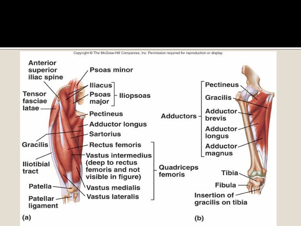
Μυς ισχίου Λαγονοψοίτης Προσαγωγοί Απαγωγοί Στροφείς Γλουτιαίοι
Ισχιοκνημιαίοι Τετρακέφαλος Ραπτικός

30
Μυς ισχίου Λαγόνιος Ψοίτης

31
Προσαγωγοί Μακρός προσαγωγός Βραχύς Ισχνός Κτενίτης Μέγας

32
Γλουτιαίοι

33
Λαγονοκνημιαία ταινία/Τείνων τη πλατεία περιτονία

34
Τετρακέφαλος

35
Απιοειδής Διπλή δράση Έσω στροφή σε κάμψη Έξω στροφή σε έκταση

36
Ισχιοκνημιαίοι Δικέφαλος Ημιτενοντώδης Ημιυμενώδης
Biceps Femoris: Long Head (Visible) Biceps femoris is a muscle of the posterior (hamstring) compartment of the thigh, and lies in the postero-lateral part of the thigh (animation) . Biceps femoris arises proximally by two ‘heads’ - termed the ‘long head’ and ‘short head’. Proximal Attachment The long head of biceps femoris (image) arises in common with the tendon of origin of semitendinosus from the supero-medial part of the ischial tuberosity. The short head has a continuous origin from the lateral lip of the linea aspera . Distal Attachment Distally, the two muscular heads fuse and give rise to a tendon, which inserts principally onto the lateral surface of the fibular head (image) . Nerve Supply The two heads of biceps femoris are innervated separately by the sciatic nerve; the long head of biceps femoris is supplied by the tibial component and the short head by the common peroneal component. Action In common with the other hamstring muscles, the action of biceps femoris is to flex the knee joint (movie) and to assist in extending the hip joint. It also laterally rotates the hip joint when the hip is extended and laterally rotates the lower leg (movie) when the knee is semi-flexed. Semimembranosus - arises from a strong membranous tendon, which gives the muscle its name. Proximal Attachment Proximally, semimembranosus arises from the supero-lateral aspect of the ischial tuberosity by a long and flattened tendon. The tendon arises from the upper lateral facet of the ischial tuberosity and continues along the lateral margin of the muscle. The fleshy muscle belly lies medial and deep to the tendon and muscle of semitendinosus and the long head of biceps femoris muscles. Distal Attachment It passes downwards and medially, with the semitendinosus tendon grooving its superficial surface. It ends at the back of the knee at a tendon, which inserts into the horizontal groove at the posterior medial corner of the medial tibial condyle (image) . Semimembranosus is attached principally to the posterior surface of the medial tibial condyle. From this insertion several expansions from the tendon pass in different directions. One of the expansions (oblique popliteal ligament) runs supero-laterally behind the knee joint reinforcing the joint capsule. Another prominent expansion passes downwards and laterally to cover the popliteus muscle. This is called the ‘popliteus fascia’. A further expansion runs antero-medially along the medial aspect of the knee and partially blends with the capsule of the knee joint. Nerve Supply Semimembranosus is innervated by the tibial component of the sciatic nerve, derived from L5, S1 and 2. Action In common with the other hamstring muscles, the action of semimembranosus is to flex the knee joint and to assist in extending the hip joint. It also medially rotates the hip joint when the hip is extended and medially rotates the lower leg (movie) when the knee is semi-flexed. Semitendinosus Proximal Attachment : arises from the supero-medial part of the ischial tuberosity of the hip bone, in common with the tendon of origin of the long head of the biceps femoris muscle. From this origin, the semitendinosus muscle runs obliquely, infero-medially behind semimembranosus. Approximately halfway down the thigh, the semitendinosus muscle gives rise to a strong, rounded tendon. Distal Attachment In the lower part of the thigh, semitendinosus (animation) and semimembranosus together form the upper medial boundary of the popliteal fossa. Distally, semitendinosus is attached to the upper part of the medial surface of the tibial shaft postero-inferior to the insertion of sartorius and gracilis. Nerve Supply Semitendinosus is innervated by the tibial component of the sciatic nerve, derived from L5, S1 and 2. Action As it is a hamstring muscle (movie) , its action is to assist in flexion of the knee and extension of the hip joint. It also medially rotates the hip joint when the hip is extended and medially rotates the lower leg when the knee is semi-flexed.
 Biceps femoris is a muscle of the posterior (hamstring) compartment of the thigh, and lies in the postero-lateral part of the thigh (animation) . Biceps femoris arises proximally by two ‘heads’ - termed the ‘long head’ and ‘short head’. Proximal Attachment The long head of biceps femoris (image) arises in common with the tendon of origin of semitendinosus from the supero-medial part of the ischial tuberosity. The short head has a continuous origin from the lateral lip of the linea aspera . Distal Attachment. Distally, the two muscular heads fuse and give rise to a tendon, which inserts principally onto the lateral surface of the fibular head (image) . Nerve Supply The two heads of biceps femoris are innervated separately by the sciatic nerve; the long head of biceps femoris is supplied by the tibial component and the short head by the common peroneal component. Action In common with the other hamstring muscles, the action of biceps femoris is to flex the knee joint (movie) and to assist in extending the hip joint. It also laterally rotates the hip joint when the hip is extended and laterally rotates the lower leg (movie) when the knee is semi-flexed. Semimembranosus - arises from a strong membranous tendon, which gives the muscle its name. Proximal Attachment Proximally, semimembranosus arises from the supero-lateral aspect of the ischial tuberosity by a long and flattened tendon. The tendon arises from the upper lateral facet of the ischial tuberosity and continues along the lateral margin of the muscle. The fleshy muscle belly lies medial and deep to the tendon and muscle of semitendinosus and the long head of biceps femoris muscles. Distal Attachment It passes downwards and medially, with the semitendinosus tendon grooving its superficial surface. It ends at the back of the knee at a tendon, which inserts into the horizontal groove at the posterior medial corner of the medial tibial condyle (image) . Semimembranosus is attached principally to the posterior surface of the medial tibial condyle. From this insertion several expansions from the tendon pass in different directions. One of the expansions (oblique popliteal ligament) runs supero-laterally behind the knee joint reinforcing the joint capsule. Another prominent expansion passes downwards and laterally to cover the popliteus muscle. This is called the ‘popliteus fascia’. A further expansion runs antero-medially along the medial aspect of the knee and partially blends with the capsule of the knee joint. Nerve Supply Semimembranosus is innervated by the tibial component of the sciatic nerve, derived from L5, S1 and 2. Action In common with the other hamstring muscles, the action of semimembranosus is to flex the knee joint and to assist in extending the hip joint. It also medially rotates the hip joint when the hip is extended and medially rotates the lower leg (movie) when the knee is semi-flexed. Semitendinosus. Proximal Attachment : arises from the supero-medial part of the ischial tuberosity of the hip bone, in common with the tendon of origin of the long head of the biceps femoris muscle. From this origin, the semitendinosus muscle runs obliquely, infero-medially behind semimembranosus. Approximately halfway down the thigh, the semitendinosus muscle gives rise to a strong, rounded tendon. Distal Attachment In the lower part of the thigh, semitendinosus (animation) and semimembranosus together form the upper medial boundary of the popliteal fossa. Distally, semitendinosus is attached to the upper part of the medial surface of the tibial shaft postero-inferior to the insertion of sartorius and gracilis. Nerve Supply. Semitendinosus is innervated by the tibial component of the sciatic nerve, derived from L5, S1 and 2. Action As it is a hamstring muscle (movie) , its action is to assist in flexion of the knee and extension of the hip joint. It also medially rotates the hip joint when the hip is extended and medially rotates the lower leg when the knee is semi-flexed.")
37
Νεύρα Μηριαίο νεύρο- λαγονοσφυικό πλέγμα ( Ο2,3,4)τετρακέφαλος), λαγονοψοίτης, κτενίτης. Νευρώνει όλους τους μυς της πρόσθιας επιφάνειας μηρού Θυροειδές νεύρο( βραχύς προσαγωγός, μακρός, ισχνός προσαγωγός) + αισθητικό κλάδο Femoral Nerve Anatomy Text The femoral nerve (image) is a branch of the lumbosacral plexus. It arises from the posterior divisions of the ventral rami of the second, third and fourth lumbar nerves. It passes infero-laterally through the substance of psoas major (animation) , behind the obturator nerve, gaining the groove between the psoas major and iliacus muscles, just below the iliac crest. It descends in the groove behind the iliac fascia. Within the abdomen, it gives off branches to iliacus, pectineus and the femoral artery. The nerve (or nerves) to pectineus arises close to the inguinal ligament, passing behind the femoral vessels to the lateral border of the muscle. The nerve enters the thigh posterior to the inguinal ligament and lateral to the femoral artery and femoral sheath. It divides into anterior and posterior divisions. It supplies all the muscles in the anterior compartment of the thigh (image) . It also gives articular branches to the hip and knee joints. A branch to the hip joint arises from the nerve to rectus femoris and branches to the knee joint arise from each of the nerves supplying the vastus muscles with a fourth branch possibly arising from the saphenous nerve Obturator Nerve The obturator nerve (image) arises from the ventral divisions of the ventral rami of the second, third and fourth lumbar nerves. It descends through psoas major, emerging from its medial border at the pelvic brim. It passes behind the common iliac vessels and descends lateral to the internal iliac vessels, along the lateral wall of the true pelvis, where it lies on the obturator internus muscle (image) . It enters the thigh through the upper part of the obturator foramen. Here it separates into anterior and posterior branches. The anterior branch descends (image) in front of obturator externus and adductor brevis and behind pectineus and adductor longus. It gives a branch to the hip joint as it enters the thigh. Branches to adductor longus, gracilis, adductor brevis and sometimes pectineus, arise as the nerve descends between the muscle layers. At the lower border of adductor longus, it contributes a branch to the subsartorial plexus. Terminally, it gives off a vascular and sometimes a cutaneous branch. When present, the cutaneous branch passes between gracilis and adductor longus to supply the skin over the lower two-thirds of the medial side of the thigh. The posterior branch penetrates and supplies the obturator externus muscle, and then descends between adductor brevis and magnus. It penetrates adductor magnus, supplying its upper part, to enter the popliteal fossa behind the popliteal artery. It supplies the posterior aspect of the knee joint and the cruciate ligaments.
 + αισθητικό κλάδο. Femoral Nerve Anatomy Text The femoral nerve (image) is a branch of the lumbosacral plexus. It arises from the posterior divisions of the ventral rami of the second, third and fourth lumbar nerves. It passes infero-laterally through the substance of psoas major (animation) , behind the obturator nerve, gaining the groove between the psoas major and iliacus muscles, just below the iliac crest. It descends in the groove behind the iliac fascia. Within the abdomen, it gives off branches to iliacus, pectineus and the femoral artery. The nerve (or nerves) to pectineus arises close to the inguinal ligament, passing behind the femoral vessels to the lateral border of the muscle. The nerve enters the thigh posterior to the inguinal ligament and lateral to the femoral artery and femoral sheath. It divides into anterior and posterior divisions. It supplies all the muscles in the anterior compartment of the thigh (image) . It also gives articular branches to the hip and knee joints. A branch to the hip joint arises from the nerve to rectus femoris and branches to the knee joint arise from each of the nerves supplying the vastus muscles with a fourth branch possibly arising from the saphenous nerve. Obturator Nerve. The obturator nerve (image) arises from the ventral divisions of the ventral rami of the second, third and fourth lumbar nerves. It descends through psoas major, emerging from its medial border at the pelvic brim. It passes behind the common iliac vessels and descends lateral to the internal iliac vessels, along the lateral wall of the true pelvis, where it lies on the obturator internus muscle (image) . It enters the thigh through the upper part of the obturator foramen. Here it separates into anterior and posterior branches. The anterior branch descends (image) in front of obturator externus and adductor brevis and behind pectineus and adductor longus. It gives a branch to the hip joint as it enters the thigh. Branches to adductor longus, gracilis, adductor brevis and sometimes pectineus, arise as the nerve descends between the muscle layers. At the lower border of adductor longus, it contributes a branch to the subsartorial plexus. Terminally, it gives off a vascular and sometimes a cutaneous branch. When present, the cutaneous branch passes between gracilis and adductor longus to supply the skin over the lower two-thirds of the medial side of the thigh. The posterior branch penetrates and supplies the obturator externus muscle, and then descends between adductor brevis and magnus. It penetrates adductor magnus, supplying its upper part, to enter the popliteal fossa behind the popliteal artery. It supplies the posterior aspect of the knee joint and the cruciate ligaments.")
38
Νεύρα Ισχιακό νεύρο Sciatic Nerve Anatomy Text The sciatic nerve (image) is the largest nerve in the body, and consists of the medially placed tibial nerve and the laterally placed common peroneal nerve. It is formed from the ventral rami of the fourth lumbar to third sacral spinal nerves and is a continuation of the upper band of the sacral plexus. It leaves the pelvis through the greater sciatic foramen, below the piriformis muscle (animation) , and descends between the greater trochanter of the femur and the ischial tuberosity. Initially deep to piriformis, it runs inferiorly and laterally posterior to the ischium, crossing over the nerve to quadratus femoris. Inferior to piriformis; it lies deep to gluteus maximus. It passes inferiorly crossing obturator internus, the gemelli and quadratus femoris. The posterior cutaneous nerve of thigh (image) and the inferior gluteal artery lie on its medial side. Descending vertically, it enters the thigh at the lower border of gluteus maximus, where it lies on the posterior surface of adductor magnus (animation) . It gives off nerves to the hamstring muscles. The nerve is crossed obliquely on its superficial aspect by the long head of biceps femoris (image) . The nerve ends at the upper aspect of the popliteal fossa by dividing into the tibial and common perineal nerves. The nerve can be represented on the back of the thigh by a line drawn from just medial to the midpoint of the line from the ischial tuberosity to the apex of greater trochanter down to the apex of popliteal fossa. It supplies articular branches to the hip joint, with muscular branches to biceps femoris, semitendinosus and semimembranosus and the ischial head of adductor magnus. The nerve to the short head of biceps is from the common peroneal division, with the other muscular branches emerging from the tibial division.
 is the largest nerve in the body, and consists of the medially placed tibial nerve and the laterally placed common peroneal nerve. It is formed from the ventral rami of the fourth lumbar to third sacral spinal nerves and is a continuation of the upper band of the sacral plexus. It leaves the pelvis through the greater sciatic foramen, below the piriformis muscle (animation) , and descends between the greater trochanter of the femur and the ischial tuberosity. Initially deep to piriformis, it runs inferiorly and laterally posterior to the ischium, crossing over the nerve to quadratus femoris. Inferior to piriformis; it lies deep to gluteus maximus. It passes inferiorly crossing obturator internus, the gemelli and quadratus femoris. The posterior cutaneous nerve of thigh (image) and the inferior gluteal artery lie on its medial side. Descending vertically, it enters the thigh at the lower border of gluteus maximus, where it lies on the posterior surface of adductor magnus (animation) . It gives off nerves to the hamstring muscles. The nerve is crossed obliquely on its superficial aspect by the long head of biceps femoris (image) . The nerve ends at the upper aspect of the popliteal fossa by dividing into the tibial and common perineal nerves. The nerve can be represented on the back of the thigh by a line drawn from just medial to the midpoint of the line from the ischial tuberosity to the apex of greater trochanter down to the apex of popliteal fossa. It supplies articular branches to the hip joint, with muscular branches to biceps femoris, semitendinosus and semimembranosus and the ischial head of adductor magnus. The nerve to the short head of biceps is from the common peroneal division, with the other muscular branches emerging from the tibial division.")
39
Femoral Nerve Innervates the: Iliopsoas Pectineus Sartorius
Rectus Femoris

40
Ψηλάφηση Που είναι το μηριαίο τρίγωνο;
Περιοχή μεταξύ του κάτω άκρου και του κορμού όρια: Βουβωνικός σύνδεσμος Μακρός προσαγωγός Ραπτικός Τι εμπεριέχει; IST/UH ΝΜΣ1

41
Μηριαίο νεύρο και μηριαία αρτηρία
Μηριαίο Τρίγωνο Βουβωνικός σύνδεσμος Μηριαίο Τρίγωνο Μηριαίο νεύρο και μηριαία αρτηρία Ραπτικός Μακρός προσαγωγός Κορυφή IST/UH ΝΜΣ1

42
Χήνειος πόδας Ραπτικός Ισχνός Ημιτενοντώδης IST/UH ΝΜΣ1

44
Την επόμενη εβδομάδα Επανάληψη…..

45
Psoas Major (Iliopsoas)
Origin Bodies and transverse processes of lumbar vertebrae Insertion Lesser trochanter of femur

46
Iliacus (Iliopsoas) Origin Iliac fossa Insertion
Psoas major tendon, onto lesser trochanter of femur

47
Pectineus Origin Pectineal line on superior ramus of pubis Insertion
Pectineal line of femur (line from lesser trochanter to linea aspera)
")
48
Sartorius Origin ASIS Insertion
Upper part of medial side of tibia (pes anserinus)
")
49
Gluteal Nerve Innervates the: Gluteus Maximus Gluteus Medius
Gluteus Minimus Tensor Fasciae Latae

50
Gluteus Maximus Origin Posterior iliac crest Posterior sacrum Coccyx
Insertion Gluteal tuberosity of femur (upper) Iliotibial tract (lower)
 Iliotibial tract (lower)")
51
Gluteus Medius Origin Gluteal surface of ilium between posterior and anterior gluteal lines Insertion Lateral surface of greater trochanter

52
Gluteus Minimus Origin
Gluteal surface of ilium between anterior and inferior gluteal lines Insertion Anterior border of greater trochanter

53
Tensor Fasciae Latae Origin Iliac crest posterior to the ASIS
Insertion Iliotibial tract into Gerdy’s tubercle

54
Sacral Nerves Innervate the six deep lateral rotators: Piriformis ER

55
Piriformis Origin Anterior surface of sacrum Insertion
Greater trochanter

56
Obturator Nerve Innervates the: Adductor Brevis Adductor Longus
Adductor Magnus Gracilis

57
Adductor Brevis Origin Inferior ramus of pubis Insertion
Pectineal line and medial lip of linea aspera

58
Adductor Longus Origin Anterior body of pubis Insertion
Medial lip of linea aspera

59
Adductor Magnus Origin Inferior ramus of pubis Ramus of ischium
Ischial tuberosity Insertion Linea aspera Adductor tubercle of femur

60
Gracilis Origin Lower half of body and inferior ramus of pubis
Insertion Upper part of medial surface of tibia (pes anserinus)
")
Παρόμοιες παρουσιάσεις