Κατέβασμα παρουσίασης
Η παρουσίαση φορτώνεται. Παρακαλείστε να περιμένετε

1
Ανατομία dsfsf Νευρικό Σύστημα dsfsf dsfsf Ελευθερία Θωμαΐδου, Pt

2
Το νευρικο συστημα

3
Μαθησιακα αποτελεσματα
Στο τέλος αυτής της ενότητας και μετά από την ανεξάρτητη μελέτη πρέπει να είστε σε θέση να: Κατανοήσετε τη βασική ανατομία και φυσιολογία του νευρικού συστήματος Αναγνωρίσετε τις διαφορές μεταξύ του Κεντρικού Νευρικού Συστήματος ( ΚΝΣ) και του Περιφερικού Νευρικού Συστήματος( ΠΝΣ) Περιγράψετε τη διαδρομή ενός ερεθίσματος από τον αισθητικό υποδοχέα μέχρι το μυ και την κινητική απάντηση.
 και του Περιφερικού Νευρικού Συστήματος( ΠΝΣ) Περιγράψετε τη διαδρομή ενός ερεθίσματος από τον αισθητικό υποδοχέα μέχρι το μυ και την κινητική απάντηση.")
4
Basic Anatomy of the Nervous System
1. Neuroglia - Supporting cells 2. Neurons – Excitable cells

5
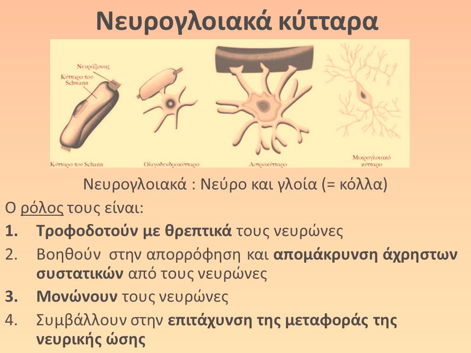
Νευρικός ιστός Ο νευρικός ιστός αποτελείται από
νευρικά κύτταρα ( νευρώνες) για τη νευρική αγωγή και νευρογλοικά κύτταρα που είναι στηρικτικά και απομονωτικά των ν.κυττάρων Τα νευρικά κύτταρα δεν πολλαπλασιάζονται και αριθμός τους παραμένει σταθερός κατά διάρκεια της ζωής….
 για τη νευρική αγωγή. και νευρογλοικά κύτταρα που είναι στηρικτικά και απομονωτικά των ν.κυττάρων. Τα νευρικά κύτταρα δεν πολλαπλασιάζονται. και αριθμός τους παραμένει σταθερός κατά. διάρκεια της ζωής….")
6
Κάθε νευρικό κύτταρο αποτελείται από:
το κυττταρικό σώμα που είναι το τροφικό κέντρο του κυττάρου Το νευρίτη ή νευράξονα που μεταφέρει τις νευρικές ώσεις Τους δενδρίτες με τις διακλαδώσεις τους Τα τελικά δενδρύλια με τα διογκωμένα άκρα τους ,τα τελικά κομβία που μεταφέρουν ερεθίσματα σε άλλα ν. κύτταρα και Τα έλυτρα

8
Τα νευρικά κύτταρα συνδέονται το ένα με το άλλο με συνάψεις
Με την απελευθέρωση νευροδιαβιβαστικών ουσιών όπως ακετυλοχολίνη, νοραδρεναλίνη, ντοπαμίνη, σεροτονίνη

9
The principle way neurons send signals over long distances is by generating and propagating an action potential or nerve impulse? an electrical signal generated / propagated along excitable cell membrane initiates many biological functions CNS neurons work in pools to integrate incoming information before sending it on

10
Το νευρικό Σύστημα Το νευρικό σύστημα στον άνθρωπο διαιρείται σε εγκεφαλονωτιαίο και αυτόνομο ή φυτικό νευρικό σύστημα Το εγκεφαλονωτιαίο νευρικό σύστημα διαιρείται σε κεντρικό ( εγκέφαλος και νωτιαίος μυελός) και περιφερικό ( εγκεφαλικά νεύρα, νωτιαία νεύρα, και νευρικά γάγγλια) Το αυτόνομο ή φυτικό νευρικό σύστημα διαιρείται σε συμπαθητικό και παρασυμπαθητικό
 και περιφερικό ( εγκεφαλικά νεύρα, νωτιαία νεύρα, και νευρικά γάγγλια) Το αυτόνομο ή φυτικό νευρικό σύστημα διαιρείται σε συμπαθητικό και παρασυμπαθητικό.")
11
The Nervous System Central nervous system (CNS)
Brain Spinal cord (SC) Peripheral nervous system (PNS) Nerves extending from brain and SC
 Peripheral nervous system (PNS) Nerves extending from brain and SC.")
12
Marieb and Hoehn- Anatomy and Physiology

13
Relationships between CNS, PNS and the environment.

14
CNS: The Brain Cerebral hemispheres Diencephalon Cerebellum Midbrain
Pons Medulla Oblongata There is a complex interaction between the cortex, cerebellum and basal ganglia, structures involved in the idea, planning, preparation and execution of movement

15
Ο εγκεφαλος Βρίσκεται μέσα στην κρανιακή κοιλότητα
Έχει βάρος γραμ στον άνδρα και γραμ στη γυναίκα Διακρίνεται σε : Τελικό με δύο ημισφάιρια Διάμεσο με τους οπτικούς θαλάμους Μέσο με το τετράδυμο και τα εγκεφαλικά σκέλη Οπίσθιο με τη γέφυρα και την παρεγκεφαλίδα Έσχατο με τον προμήκη

16
Ο εγκεφαλος Για περιγραφικούς λόγους διαιρείται σε : 2 ημισφαίρια
Για περιγραφικούς λόγους διαιρείται σε : 2 ημισφαίρια Το στέλεχος Την παρεγκεφαλίδα

17
CNS: The Brain – can you name these parts?

18
CNS: The Brain

19
Τα εγκεφαλικά ημισφαίρια αποτελούνται από ένα εξωτερικό τμήμα, φλοιός ( φαιά ουσία) που περιέχει σώματα κυττάρων και ένα εσωτερικό τμήμα ( λευκή ουσία) που αποτελείται από νευράξονες που σχηματίζουν οδούς ή δεμάτια και από τις κοιλίες, που είναι χώροι γεμάτοι εγκεφαλονωτιαίο υγρό. Ο χιτώνας του κάθε ημισφαιρίου αποτελείται: Κεντρική μοίρα ή νήσος του Reil Ο μετωπιαίος λοβός Ο βραγματικός λοβός Ο κροταφικός λοβός και Ο ινιακός λοβός

20
Ο χιτώνας των ημισφαιρίων διασχίζεται από αύλακες , μεταξύ των οποίων σχηματίζονται έλικες
Αύλακες * η κεντρική ή Ρολάνδειος ( μετωπιαίου – βρεγματικού λοβού) *ή πλάγια σχισμή του Silvius (μετωπιαίου- βρεγματικού κροταφικού) * βρεγματοΐνιακή σχισμή( βρεγματικού- ινιακού) Έλικες (πτυχές)*πρόσθια κεντρική έλικα * η έλικα του Broca , αριστερά στους δεξιόχειρες , το κέντρο του λόγου
 *ή πλάγια σχισμή του Silvius (μετωπιαίου- βρεγματικού- κροταφικού) * βρεγματοΐνιακή σχισμή( βρεγματικού- ινιακού) Έλικες (πτυχές)*πρόσθια κεντρική έλικα. * η έλικα του Broca , αριστερά στους δεξιόχειρες , το κέντρο του λόγου.")
21
Function of the Major Brain Regions
Cerebral hemispheres Interpret sensory input Controls voluntary muscle activity Intellectual and emotional processing Diencephalon Thalamus - Relay station of sensory and motor impulses to and from the cortex, Hypothalamus – Regulation centre eg body temperature Brain Stem = midbrain, pons, medulla Conduction pathway between higher, lower brain centres and the spinal cord Site of many of the cranial nerves Cerebellum Processing centre controlling balance and. co-ordination

23
Παρεγκεφαλιδα ( cerebellum)
Η Παρεγκεφαλίδα είναι κεντρικό όργανο το οποίο ρυθμίζει και συντονίζει συνειδητές και αυτόματες κινήσεις του σώματος και εξασφαλίζει την ισορροπία του σε στάση και κίνηση. Αποτελείται από τρεις λοβούς, ένα μέσο( το σκώληκα), και δύο πλάγιους( τα ημισφαίρια)
, και δύο πλάγιους( τα ημισφαίρια)")
24
μηνιγγες Πρόκειται για χιτώνες συνδετικού ιστού που περιβάλλουν, προστατεύουν και συγκροτούν τον εγκέφαλο και το νωτιαίο μυελό: Η σκληρή μήνιγγα είναι ο παχύτετρος εξωτερικός χιτώνας Η αραχνοειδής η χοριοειδής συμφύεται με το εγκέφαλο και το νωτιαίο μυελών Υπαραχνοειδής χώρος που περιέχει το ΕΝΥ

25
Νευρικες οδοι του εγκεφαλου- συζυγιες
Η πυραμιδική ( κινητική ) οδός Η αισθητική οδός( εν τω βάθει αισθητικότητα) Η οπτική οδός Η κοχλιακή οδός ( ακουστική κυρίως) Η αιθουσαία οδός Η οσφρητική οδός
 οδός. Η αισθητική οδός( εν τω βάθει αισθητικότητα) Η οπτική οδός. Η κοχλιακή οδός ( ακουστική κυρίως) Η αιθουσαία οδός. Η οσφρητική οδός.")
26
Εγκεφαλικα νευρα- συζυγιες
1. ΟΣΦΡΗΤΙΚΟ( Α) 2.ΟΠΤΙΚΟ(Α) 3.ΚΟΙΝΟ ΚΟΙΝΗΤΙΚΟ(Κ) 4.ΤΡΟΧΙΛΙΑΚΟ(Κ) 5.ΤΡΙΔΥΜΟ(Μ) 6.ΑΠΑΓΩΓΟ(Κ) 7.ΠΡΟΣΩΠΙΚΟ(Μ) 8.ΣΤΑΤΙΚΟΑΚΟΥΣΤΙΚΟ(Α) 9.ΓΛΩΣΟΦΑΡΥΓΓΙΚΟ(Μ) 10.ΠΝΕΥΜΟΝΟΓΑΣΤΡΙΚΟ( Μ) 11.ΠΑΡΑΠΛΗΡΩΜΑΤΙΚΟ( Κ) 12.ΥΠΟΓΛΩΣΣΙΟ(Κ)
 2.ΟΠΤΙΚΟ(Α) 3.ΚΟΙΝΟ ΚΟΙΝΗΤΙΚΟ(Κ) 4.ΤΡΟΧΙΛΙΑΚΟ(Κ) 5.ΤΡΙΔΥΜΟ(Μ) 6.ΑΠΑΓΩΓΟ(Κ) 7.ΠΡΟΣΩΠΙΚΟ(Μ) 8.ΣΤΑΤΙΚΟΑΚΟΥΣΤΙΚΟ(Α) 9.ΓΛΩΣΟΦΑΡΥΓΓΙΚΟ(Μ) 10.ΠΝΕΥΜΟΝΟΓΑΣΤΡΙΚΟ( Μ) 11.ΠΑΡΑΠΛΗΡΩΜΑΤΙΚΟ( Κ) 12.ΥΠΟΓΛΩΣΣΙΟ(Κ)")
27
Νωτιαιοσ Μυελος Ο νωτιαίος μυελός είναι ένα λευκό κυλινδρικό νευρικό όργανο Το άνω άκρο του είναι συνέχεια του προμήκη και το κάτω άκρο του τελειώνει στο μυελικό κώνο και στο τελικό νημάτιο Ο ΝΜ εμφανίζει τρεις μοίρες την αυχενική, τη θωρακική και την οσφυΐκή από τις οποίες εκφύονται ρίζες και νωτιαία νεύρα Ο ΝΜ δεν εκτείνεται σε όλο το μήκος του σπονδυλικού σωλήνα( έως Ο2 σπόνδυλο) τα οσφυικά και ιερά νεύρα φέρονται λοξά προς τα κάτω και σχηματίζουν την ιππουρίδα
 τα οσφυικά και ιερά νεύρα φέρονται λοξά προς τα κάτω και σχηματίζουν την ιππουρίδα.")
28
Νωτιαιος μυελος Περιέχεται στα 2/3 της ΣΣ
Νωτιαιος μυελος Περιέχεται στα 2/3 της ΣΣ Έχει κυλινδρική μορφή, και στη διατομή έχει κυκλικό σχήμα με ένα κεντρικό αυλό Εκτείνεται από το ινιακό τρήμα μέχρι το επίπεδο Ο1, Ο2 περίπου

29
Central Nervous System (CNS): Spinal Cord (SC)
Ascending tracts Sensory information to brain Descending tracts Information from brain to appropriate motor neurons Spinal reflexes Pain and Temperature Detection Receptors are free nerve endings found in most body tissue (although more densely packed in some than others) The primary afferents enter the spinal cord, synapse and then the second order neurones decussate (cross) within one or two levels of entering They then ascend the entire cord and brainstem in the Spinothalamic Tract (STT) As with the DCML tract, the STT is somatotopically organised with those afferents from the LL/trunk more laterally and UL more medially From the brainstem, the neurones continue to project and finally synapse in the thalamus From the thalamus neurones then project to the SI Discriminatory Touch and Pressure: Receptors are wide spread general sensory receptors that may either be encapsulated (e.g. Meissner’s or Pacinian corpuscles, Ruffini endings, muscle spindles, golgi tendon organs) or uncapsulated free nerve endings (e.g. Merkel discs, hair follicle receptors) Generally the RFs for these receptors is small and there is limited convergence as they travel up the spinal cord The ascending pathway involved is the Dorsal Column Medial Lemniscal System (DCML) Proprioception: Conscious proprioception is mediated via projections along the DCML Unconscious proprioception is mediated by projections along the Spinocerebellar tract (SCBT) The role of this unconscious proprioception is to monitor and modify mvts DCML System: First order sensory neurones enter the spinal cord via the dorsal horn (from both cutaneous (touch) and proprioceptive receptors) Those neurones that ascend, do not synapse in the cord, but travel ipsilaterally (same side as the afferents enter the cord) until the afferents reach the medulla In the medulla, they synapse to second order neurone, decussate and travel to the thalamus There they synapse again and travel, via the internal capsule, to the primary somatosensory cortex Primary Somatosensory Cortex (SI): This is divided into a number of regions (1, 2, 3a, 3b) that are responsible for receiving inputs predominately from different types of receptors: Area 3b – primarily cutaneous receptors Area 3a – primarily proprioceptive information from muscles Area 2 – primarily input from joints and other deep structures Area 1 – primarily from rapidly adapting cutaneous mechanoreceptors Although there is overlap in receptive field within the SI cortex, being aware that different parts of the cortex receive from different types of receptors, can help explain why, for example stroke patients may have deficit in one aspect of the somatosensory system more than others (depending on where their lesion occurred) Secondary somatosensory cortex (SII) From the SI, projections occur to the SII in the posterior parietal cortex Again this is somatotopically organised Whereas the role of the SI is to facilitate the awareness/detection of sensory information, higher order processing in the SII puts meaning to this awareness Lesions in this region lead to difficulty in making sense of information, e.g. neglect (with loss of postural schema), dyspraxias Pyrmidal = corticospinal and corticobulbar connect to virtually all mm of the body by monosynaptic connections The Corticospinal Tract Most fibres originate in the motor cortex and are then direct excitatory projections to either the motor neurones (monosynaptic) or to interneurones in the spinal cord Most fibres (approx 75%) decussate in the medulla (leading to contralateral innervation) – the others remain ipsilateral Most of the innervation from the CST goes to motor neurones controlling fine (fractionated) distal hand function Lesser amounts of innervation go to the trunk and proximal limb joints The CST develops with age – expands in density through childhood Rubrospinal from red N Vestibulospinal from vestibular N role in control of neck mm The Vestibulospinal tract: Originates from the vestibular nuclei in the brainstem These have much interconnectivity with other CNS regions and thus are able to project integrated information in respect to posture and balance The resultant output is largely postural/extensor activity of trunk and limbs Output may be either anticipatory (feedforward) or reactive (feedback) Retculospinal from RF The Reticulospinal Tract Originates in the reticular formation and projects to interneurones of the spinal cord Involved in both feedforward and feedback control of postural control (again axial/limb mm activation – both flexor and extensor) However, also involved in the regulation of tone Within the spinal cord, the majority of descending neuronal projections will synapse with interneurones It is only a number of descending neurones from the CST that synapse directly with motor neurones At the interneurones, descending neurones come together with those from the periphery, allowing for a further level of modification in respect to motor output From the anterior horn, motor neurones will exit, projecting to muscle fibres = motor unit Each muscle fibre receives innervation from just one motor neurone, but each motor neurone may innervate many muscle fibres (will depend on muscle size and specificity of activation) Motor neurones of the same motor pool are topographically mapped in the anterior horn
 The primary afferents enter the spinal cord, synapse and then the second order neurones decussate (cross) within one or two levels of entering. They then ascend the entire cord and brainstem in the Spinothalamic Tract (STT) As with the DCML tract, the STT is somatotopically organised with those afferents from the LL/trunk more laterally and UL more medially. From the brainstem, the neurones continue to project and finally synapse in the thalamus. From the thalamus neurones then project to the SI. Discriminatory Touch and Pressure: Receptors are wide spread general sensory receptors that may either be encapsulated (e.g. Meissner’s or Pacinian corpuscles, Ruffini endings, muscle spindles, golgi tendon organs) or uncapsulated free nerve endings (e.g. Merkel discs, hair follicle receptors) Generally the RFs for these receptors is small and there is limited convergence as they travel up the spinal cord. The ascending pathway involved is the Dorsal Column Medial Lemniscal System (DCML) Proprioception: Conscious proprioception is mediated via projections along the DCML. Unconscious proprioception is mediated by projections along the Spinocerebellar tract (SCBT) The role of this unconscious proprioception is to monitor and modify mvts. DCML System: First order sensory neurones enter the spinal cord via the dorsal horn (from both cutaneous (touch) and proprioceptive receptors) Those neurones that ascend, do not synapse in the cord, but travel ipsilaterally (same side as the afferents enter the cord) until the afferents reach the medulla. In the medulla, they synapse to second order neurone, decussate and travel to the thalamus. There they synapse again and travel, via the internal capsule, to the primary somatosensory cortex. Primary Somatosensory Cortex (SI): This is divided into a number of regions (1, 2, 3a, 3b) that are responsible for receiving inputs predominately from different types of receptors: Area 3b – primarily cutaneous receptors. Area 3a – primarily proprioceptive information from muscles. Area 2 – primarily input from joints and other deep structures. Area 1 – primarily from rapidly adapting cutaneous mechanoreceptors. Although there is overlap in receptive field within the SI cortex, being aware that different parts of the cortex receive from different types of receptors, can help explain why, for example stroke patients may have deficit in one aspect of the somatosensory system more than others (depending on where their lesion occurred) Secondary somatosensory cortex (SII) From the SI, projections occur to the SII in the posterior parietal cortex. Again this is somatotopically organised. Whereas the role of the SI is to facilitate the awareness/detection of sensory information, higher order processing in the SII puts meaning to this awareness. Lesions in this region lead to difficulty in making sense of information, e.g. neglect (with loss of postural schema), dyspraxias. Pyrmidal = corticospinal and corticobulbar. connect to virtually all mm of the body by monosynaptic connections. The Corticospinal Tract. Most fibres originate in the motor cortex and are then direct excitatory projections to either the motor neurones (monosynaptic) or to interneurones in the spinal cord. Most fibres (approx 75%) decussate in the medulla (leading to contralateral innervation) – the others remain ipsilateral. Most of the innervation from the CST goes to motor neurones controlling fine (fractionated) distal hand function. Lesser amounts of innervation go to the trunk and proximal limb joints. The CST develops with age – expands in density through childhood. Rubrospinal from red N. Vestibulospinal from vestibular N. role in control of neck mm. The Vestibulospinal tract: Originates from the vestibular nuclei in the brainstem. These have much interconnectivity with other CNS regions and thus are able to project integrated information in respect to posture and balance. The resultant output is largely postural/extensor activity of trunk and limbs. Output may be either anticipatory (feedforward) or reactive (feedback) Retculospinal from RF. The Reticulospinal Tract. Originates in the reticular formation and projects to interneurones of the spinal cord. Involved in both feedforward and feedback control of postural control (again axial/limb mm activation – both flexor and extensor) However, also involved in the regulation of tone. Within the spinal cord, the majority of descending neuronal projections will synapse with interneurones. It is only a number of descending neurones from the CST that synapse directly with motor neurones. At the interneurones, descending neurones come together with those from the periphery, allowing for a further level of modification in respect to motor output. From the anterior horn, motor neurones will exit, projecting to muscle fibres = motor unit. Each muscle fibre receives innervation from just one motor neurone, but each motor neurone may innervate many muscle fibres (will depend on muscle size and specificity of activation) Motor neurones of the same motor pool are topographically mapped in the anterior horn.")
30
Central Nervous System (CNS): Spinal Cord (SC)
: Spinal Cord (SC)")
31
Νωτιαιοσ μυελος Στην εγκάρσια διατομή του ΝΜ αποκαλύπτεται :
Η φαιά ουσία που καταλαμβάνει το κέντρο και Τη λευκή που βρίσκεται γύρω από τη φαιή Κάθε φαιή στήλη αποτελείται από το πρόσθιο διογκωμένο κέρας , το οπίσθιο( λεπτότερο) και τη μεσοκεράτια ζώνη Τα πρόσθια κέρατα περιλαμβάνουν κινητικά κύτταρα, οι νευρίτες των οποίων πορεύονται στο σχηματισμό των πρόσθιων ( κινητικών ) ριζών των νωτιαίων νεύρων Τα λεπτότερα και επιμηκέστερα οπίσθια κέρατα αποτελούνται από αισθητικά κύτταρα, τα οποία υποδέχονται κεντρομόλες ίνες από τις οπίσθιες ίνες από τις οπίσθιες ρίζες των νωτιαίων νεύρων
 και τη μεσοκεράτια ζώνη. Τα πρόσθια κέρατα περιλαμβάνουν κινητικά κύτταρα, οι νευρίτες των οποίων πορεύονται στο σχηματισμό των πρόσθιων ( κινητικών ) ριζών των νωτιαίων νεύρων. Τα λεπτότερα και επιμηκέστερα οπίσθια κέρατα αποτελούνται από αισθητικά κύτταρα, τα οποία υποδέχονται κεντρομόλες ίνες από τις οπίσθιες ίνες από τις οπίσθιες ρίζες των νωτιαίων νεύρων.")
32
Νωτιαιος Μυελος Στην εξωτερική του επιφάνεια παρουσιάζει δύο ογκώματα το αυχενικό ( κάτω από των χιασμό των πυραμίδων στον προμήκη) που τελειώνει στο ύψος του 2ου θωρακικού και το οσφυΐκό που αρχίζει στο ύψος του 10ου θωρακικού και τελειώνει στο μυελικό κώνο Η εξωτερική του επιφάνεια φέρει αύλακες Δια μέσου της λευκής ουσίας του ΝΜ ανεβαίνουν ( ανιούσες) και κατεβαίνουν ( κατιούσες) νευρικές οδοί.
 που τελειώνει στο ύψος του 2ου θωρακικού και το οσφυΐκό που αρχίζει στο ύψος του 10ου θωρακικού και τελειώνει στο μυελικό κώνο. Η εξωτερική του επιφάνεια φέρει αύλακες. Δια μέσου της λευκής ουσίας του ΝΜ ανεβαίνουν ( ανιούσες) και κατεβαίνουν ( κατιούσες) νευρικές οδοί.")
33
Η πυραμιδική οδός( κινητική)
Η αισθητική οδός( εν τω βαθει αισθητικότητα) Οι νωταιο- παρεγκεφαλιδικές οδοί( πρόσθια και οπίσθια)
 Οι νωταιο- παρεγκεφαλιδικές οδοί( πρόσθια και οπίσθια)")
34
Peripheral Nervous System (PNS)
sensory, motor and autonomic neurons Connections to and from the SC Connections within the SC

35
Sensory information When considering movement where might the sensory information come from? What information will be provided?

36
Sensory information When considering movement where might the sensory information come from? What information will be provided? Touch Pressure Temperature Pain Proprioception

37
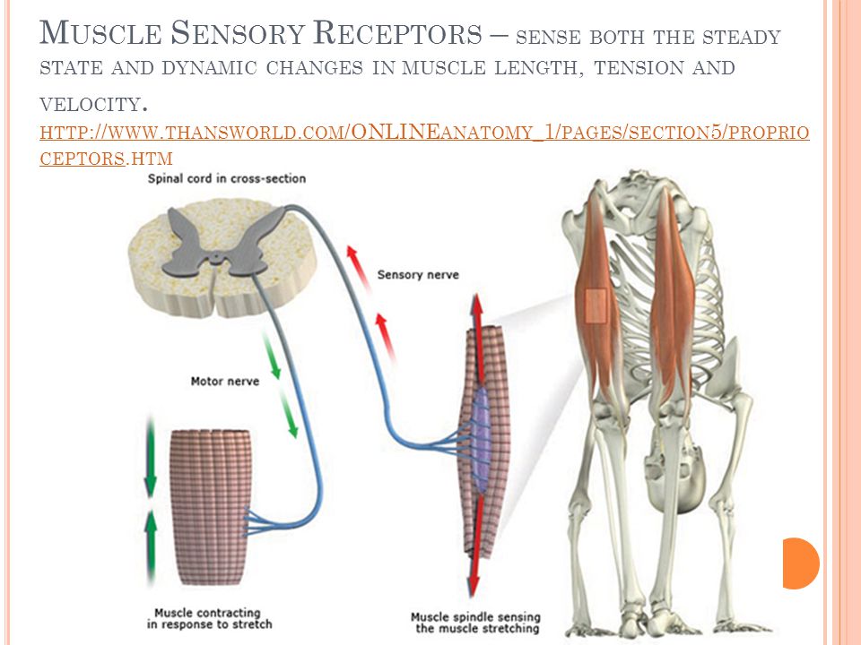
Proprioception - Muscle Sensory Receptors
Information is provided about the current position of the limb and the length and velocity of its muscles Muscle spindles Golgi Tendon Organs

38
Muscle Sensory Receptors – sense both the steady state and dynamic changes in muscle length, tension and velocity.
39
Journey a nerve impulse may take from sensory receptor to muscle
CNS Environment Task Brain SC tracts Reflex action Afferent nerves Efferent nerves PNS Motor response Muscle contraction Sensory receptors Movement

40
Journey a nerve impulse may take from CNS to muscle
Where the body is Decide on strategy Association areas of the cortex Past experience Basal ganglia Decide how to move Motor cortex, cerebellum Instructions Brainstem , spinal cord Movement & Postural adjustment

Παρόμοιες παρουσιάσεις


>")


