Κατέβασμα παρουσίασης
Η παρουσίαση φορτώνεται. Παρακαλείστε να περιμένετε

1
Σκωληκοειδιτιδα

2
σκωληκοειδης αποφυση ΣΚΩΛΗΚΟΕΙΔΗΣ ΑΠΟΦΥΣΗ
Έχει δικό της μεσεντέριο (μεσεντερίδιο) Αιματώνεται από τελική αρτηρία, κλάδο της ειλεοκολικής αρτηρίας Η φλεβική της κυκλοφορία είναι μέσω του ειλεοκολικού φλεβ.συστ. πυλαία Η λεμφική αποχέτευση γίνεται μέσω των ειλεοκολικών λεμφαδένων που ακολουθουν την πορεία της ανω μεσεντέριας αρτηρίας Η σκωληκοειδής στο βλεννογόνο της έχει απλούς σωληνοειδείς αδένες (1). Διακρίνονται ακόμη το καλυπτήριο επιθήλιο (βέλη), το οποίο είναι μονόστιβο κυλινδρικό (βέλη) και λεμφοζίδια (2) στον βλεννογόνο της. (χρώση αιματοξυλίνη-εωσίνη, μεγέθυνση Χ50)
 Αιματώνεται από τελική αρτηρία, κλάδο της ειλεοκολικής αρτηρίας. Η φλεβική της κυκλοφορία είναι μέσω του ειλεοκολικού φλεβ.συστ. πυλαία. Η λεμφική αποχέτευση γίνεται μέσω των ειλεοκολικών λεμφαδένων που ακολουθουν την πορεία της ανω μεσεντέριας αρτηρίας. Η σκωληκοειδής στο βλεννογόνο της έχει απλούς σωληνοειδείς αδένες (1). Διακρίνονται ακόμη το καλυπτήριο επιθήλιο (βέλη), το οποίο είναι μονόστιβο κυλινδρικό (βέλη) και λεμφοζίδια (2) στον βλεννογόνο της. (χρώση αιματοξυλίνη-εωσίνη, μεγέθυνση Χ50)")
3
σκωληκοειδης αποφυση

4
Σκωληκοειδιτιδα Επιπολασμός (ΗΠΑ):
10 περιπτώσεις ανα γενικού πλυθησμού Στα παιδιά: 4 περιπτώσεις ανα παιδιά

5
Σκωληκοειδιτιδα Σε σχέση με το φύλο: 2♂:1♀
Τη στιγμή της διάγνωσης το ποσοστό διάτρησης είναι 20-35%. Σε νεαρότερες ηλικίες το ποσοστό αυτό αυξάνεται σημαντικά σε 50-85%.

6
Σκωληκοειδιτιδα Κλινική εικόνα
Τυπικός κολικός πόνος περιομφαλικά ή και στο επιγάστριο Ναυτία, εμετό, ανορεξία, πυρετική κίνηση Σημέιο McBurney, σημείο rebound, σημείο Rovsing, σημείο ψοίτη Λευκοκυττάρωση, CRP

7
Σκωληκοειδιτιδα Στάδια: Σπάνιοι τύποι: πρώιμο στάδιο σκωληκοειδίτιδας
Διαπυούσα σκωληκοειδίτιδα Γαγγραινώδης σκωληκοειδίτιδα Διατρηθείσα σκωληκοειδίτιδα Φλεγμένουσα σκωληκοειδίτιδα (plastron) Σπάνιοι τύποι: Spontaneously resolving appendicitis Recurrent appendicitis Chronic appendicitis
 Σπάνιοι τύποι: Spontaneously resolving appendicitis. Recurrent appendicitis. Chronic appendicitis.")
8
Σκωληκοειδιτιδα Υπάρχουν αντικρουόμενες απόψεις για το πότε πρέπει να ζητείτε απεικονιστική μέθοδος στην οξεία σκωληκοειδίτιδα Πριν το 1980 η απεικονιστική μέθοδος που χρησιμοποιούσαν ήταν απλή ακτινογραφία ή και βαριούχο υποκλεισμό. Partial appendiceal intussusception. The appendix is incompletely filled (solid arrow) and partially intussuscepting into the cecal caput (open arrow).
 and partially intussuscepting into the cecal caput (open arrow).")
9
Από το 1986 χρησιμοποιείται το υπερηχογράφημα και μετά το 1990 η αξονική τομογραφία, επειδή μπορούσε να δείξει την σκωληκοειδή απόφυση, το περιβάλλων λίπος και το έντερο. Έχει υποστηριχθεί ότι με τη χρήση CT μειώθηκαν κατά 21% οι αρνητικές σκωλικοειδεκτομές Schuler JG, Shortsleeve MJ, Goldenson RS, et al. Is there a role for abdominal computed tomographic scans in appendicitis? Arch Surg. 1998;133: ; discussion 377.

10
Η ευαισθησία της CT και του US έχει υποστηριχθεί ότι είναι παρόμοιες.
Η ευαισθησία του CT κυμαίνεται μεταξύ 85%-99% και του US μεταξύ 74% to 99% Horton MD, Counter SF, Florence MG, Hart MJ. A prospective trial of computed tomography and ultrasonography for diagnosing appendicitis in the atypical patient. Am J Surg. 2000;179: Lowe LH, Penney MW, Stein SM, et al. Unenhanced limited CT of the abdomen in the diagnosis of appendicitis in children: Comparison with sonography. AJR Am J Roentgenol. 2001;176:31-35.

11
Ευρhματα στο US Πεπαχυσμένο τοίχωμα >3mm Διάμετρος > 6ή 7mm Μη συμπιέσιμη σκωληκοειδής απόφυση Κοπρόλιθο Περιφεριακή αιμάτωση Ηχογενές μεσεντέριο Ελεύθερο υγρό Απόστημα

12
Ultrasound scanning technique
Ultrasound scanning technique. Linear ray ultrasound probe using compression technique is used to examine the RLQ of the abdomen Normal bowel. (A) Ultrasound examination of the RLQ demonstrating 1-cm loop of fluid-filled bowel (arrow). (B) With compression and normal peristalsis, the diameter of this normal loop of bowel decreases to 5 mm (arrow).
 Ultrasound examination of the RLQ demonstrating 1-cm loop of fluid-filled bowel (arrow). (B) With compression and normal peristalsis, the diameter of this normal loop of bowel decreases to 5 mm (arrow).")
13
Acute appendicitis. Ultrasound of the RLQ of the abdomen demonstrating blind-ended tubular structure (open arrows) corresponding to acutely inflamed appendix. Note the distended lumen [L], the echogenic surrounding mesentery [M], and the echogenic structure with acoustic shadow (arrow) at the base of the appendix corresponding to an appendolith. Acute appendicitis. Transverse color-flow ultrasound of the RLQ of the abdomen demonstrates increased vascularity (arrow) in a fluid-filled structure corresponding to acute appendicitis. External iliac artery [A] is identified.
![Acute appendicitis. Ultrasound of the RLQ of the abdomen demonstrating blind-ended tubular structure (open arrows) corresponding to acutely inflamed appendix. Note the distended lumen [L], the echogenic surrounding mesentery [M], and the echogenic structure with acoustic shadow (arrow) at the base of the appendix corresponding to an appendolith.](http://slideplayer.gr/slide/2848745/10/images/13/Acute+appendicitis.+Ultrasound+of+the+RLQ+of+the+abdomen+demonstrating+blind-ended+tubular+structure+%28open+arrows%29+corresponding+to+acutely+inflamed+appendix.+Note+the+distended+lumen+%5BL%5D%2C+the+echogenic+surrounding+mesentery+%5BM%5D%2C+and+the+echogenic+structure+with+acoustic+shadow+%28arrow%29+at+the+base+of+the+appendix+corresponding+to+an+appendolith..jpg "Acute appendicitis. Transverse color-flow ultrasound of the RLQ of the abdomen demonstrates increased vascularity (arrow) in a fluid-filled structure corresponding to acute appendicitis. External iliac artery [A] is identified.")
14
Acute appendicitis. Ultrasound demonstrating a blind-ended tubular structure with fluid-filled lumen [L]. Also note there is a small amount of fluid [M] surrounding the tip of the appendix. Suppurative appendicitis; transverse view, Circumferential colors are observed in the wall of the inflamed appendix (arrows), a strong indicator of acute appendicitis.
![Acute appendicitis. Ultrasound demonstrating a blind-ended tubular structure with fluid-filled lumen [L]. Also note there is a small amount of fluid [M] surrounding the tip of the appendix.](http://slideplayer.gr/slide/2848745/10/images/14/Acute+appendicitis.+Ultrasound+demonstrating+a+blind-ended+tubular+structure+with+fluid-filled+lumen+%5BL%5D.+Also+note+there+is+a+small+amount+of+fluid+%5BM%5D+surrounding+the+tip+of+the+appendix..jpg "Suppurative appendicitis; transverse view, Circumferential colors are observed in the wall of the inflamed appendix (arrows), a strong indicator of acute appendicitis.")
15
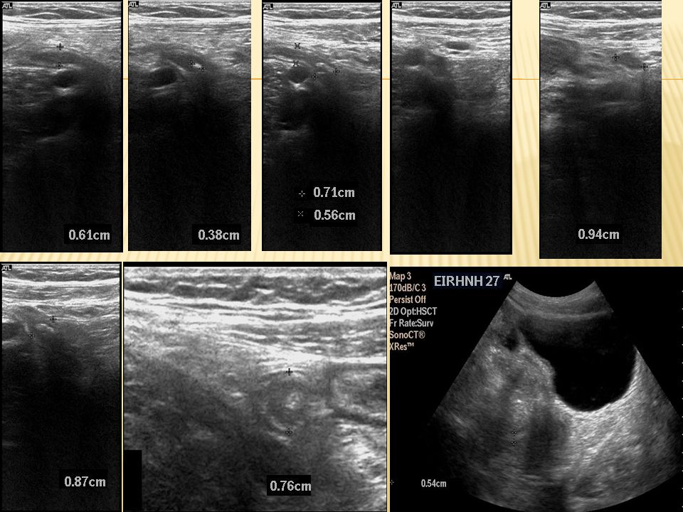
Suppurative appendicitis in a 15-year-old boy (same patient )
An inflamed appendix with appendicoliths in the lumen (arrow) is demonstrated. Suppurative appendicitis in a 15-year-old boy; longitudinal ultrasonogram. An aperistaltic, noncompressible, blind-ended, fluid-filled, tubular appendiceal structure is shown, and distinct wall layers (arrows) arising from the base of the cecum are observed. Normal appendix; longitudinal ultrasonogram. A compressible tubular appendiceal structure with an outer diameter of less than 6 mm (arrows) is shown. A = iliac artery; V = iliac vein.
 is demonstrated. Suppurative appendicitis in a 15-year-old boy; longitudinal ultrasonogram. An aperistaltic, noncompressible, blind-ended, fluid-filled, tubular appendiceal structure is shown, and distinct wall layers (arrows) arising from the base of the cecum are observed. Normal appendix; longitudinal ultrasonogram. A compressible tubular appendiceal structure with an outer diameter of less than 6 mm (arrows) is shown. A = iliac artery; V = iliac vein.")
16
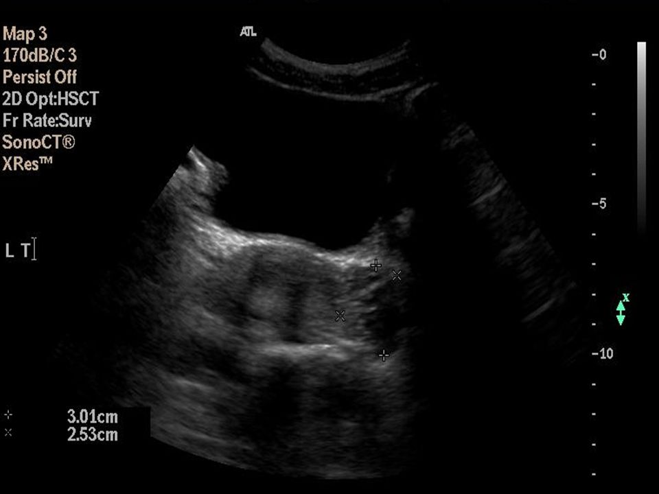
(Same patient) Gangrenous appendicitis; longitudinal ultrasonogram. A markedly distended appendix (arrows), loss of mucosa and submucosal layers, and prominent echogenic pericecal fat are shown. Phlegmonous appendicitis; oblique-axial ultrasonogram. A pericecal fluid collection, which is walled off by small-bowel loops (arrowheads) is shown, and an appendicolith with an acoustic shadow (arrow) is observed
, loss of mucosa and submucosal layers, and prominent echogenic pericecal fat are shown. Phlegmonous appendicitis; oblique-axial ultrasonogram. A pericecal fluid collection, which is walled off by small-bowel loops (arrowheads) is shown, and an appendicolith with an acoustic shadow (arrow) is observed.")
17
Perforated appendix; longitudinal ultrasonogram
Perforated appendix; longitudinal ultrasonogram. A defect on the tip (large arrow, right side) of the enlarged appendix (short arrows, left side) is observed. c = cecum Periappendiceal abscess formation; oblique-axial ultrasonogram. A thick-walled, complex, hypoechoic mass adjacent to the cecum (arrows) is shown. The inflamed appendix was not visualized
 of the enlarged appendix (short arrows, left side) is observed. c = cecum. Periappendiceal abscess formation; oblique-axial ultrasonogram. A thick-walled, complex, hypoechoic mass adjacent to the cecum (arrows) is shown. The inflamed appendix was not visualized.")
Παρόμοιες παρουσιάσεις


 Όραση Μαρία Κουτρομάνου. Structure of the Eye: Iris The iris is similar to the diaphragm in a camera Your iris widens in dim light and.>")

