Κατέβασμα παρουσίασης
Η παρουσίαση φορτώνεται. Παρακαλείστε να περιμένετε

1
Γεννητικό σύστημα θήλεος Τράχηλος

2
Here is a normal cervix with a smooth, glistening mucosal surface
Here is a normal cervix with a smooth, glistening mucosal surface. There is a small rim of vaginal cuff from this hysterectomy specimen. The cervical os is small and round, typical for a nulliparous woman. The os will have a fish-mouth shape after one or more pregnancies.

3
Schematic Representation of Normal Stratified Squamous EpitheliumThe process of epithelial differentiation and cell turnover (in which basal stem cells give rise to daughter cells that ultimately reach the surface of the epithelium as mature keratinocytes) takes several weeks. The process begins when the cells in the basal layer divide along the basement membrane spreading horizontally. Some progeny cells begin to migrate upward. Once no longer attached to the basement membrane, these cells exit the cell cycle, stop dividing, and begin to differentiate. Cells reaching the superficial zone die and are shed from the surface [Sompayrac, 2002]. Hence, in normal squamous epithelium the maturation and differentiation of cells throughout the epithelial thickness is carried out in an orderly manner (Figure 20).
..")
4
This is normal cervical non-keratinizing squamous epithelium
This is normal cervical non-keratinizing squamous epithelium. The squamous cells show maturation from basal layer to surface.

5
Micro anatomy of the uterine cervix: 1= Nulliparous, 2= Multiparous (A: external os, pink area = non keratinized squamous epithelium, purple area = glandular epithelium composed of one layer of mucin secreting and ciliated cells).
.")
6
Structure of the ectocervix: CT=connective tissue, BM=basement membrane, L1=basal cells (1 layer), L2=parabasal cells (2 layers), L3=intermediate cells (around 8 layers), L4=superficial cells (5 or 6 layers) and L5=exfoliating cells.
, L2=parabasal cells (2 layers), L3=intermediate cells (around 8 layers), L4=superficial cells (5 or 6 layers) and L5=exfoliating cells.")
7
Distribution of the endocervical glands
Distribution of the endocervical glands. (A=External os, B=Isthmus and C= Limits of the glandular field).
.")
8
epithelium composed of one layer of mucin secreting cells with few ciliated cells (+).
.")
9
epithelium composed of one layer of mucin secreting cells with few reserve cells (arrow).
.")
10
The Transformation Zone
In most cases, cellular changes leading to precancerous/cancerous lesions of the cervix take place in the transformation zone, the region between the original and current squamocolumnar junctions (Figure 25). This is where the process of squamous metaplasia occurs – the gradual replacement of the columnar epithelium of the endocervix by squamous epithelium. Immature metaplastic epithelium is actively proliferating at this junctional site, and is particularly vulnerable to transformation to precancerous and cancerous lesions [Koss, 1999]. Furthermore, unlike the stratified squamous epithelium which consists of multiple cell layers, the transformation zone is composed of only a few layers of cells so that it is more susceptible to tears/microtrauma through which HPV can be introduced and infect basal cells. In addition to the cervix, squamocolumnar junctions exist at other body sites, such as where the anal epithelium meets the rectal epithelium, and certain sites in the upper airway. These are also very susceptible to HPV infection.
. This is where the process of squamous metaplasia occurs – the gradual replacement of the columnar epithelium of the endocervix by squamous epithelium. Immature metaplastic epithelium is actively proliferating at this junctional site, and is particularly vulnerable to transformation to precancerous and cancerous lesions [Koss, 1999]. Furthermore, unlike the stratified squamous epithelium which consists of multiple cell layers, the transformation zone is composed of only a few layers of cells so that it is more susceptible to tears/microtrauma through which HPV can be introduced and infect basal cells. In addition to the cervix, squamocolumnar junctions exist at other body sites, such as where the anal epithelium meets the rectal epithelium, and certain sites in the upper airway. These are also very susceptible to HPV infection.")
11
Φλεγμονές τραχήλου Ενδογενή αερόβια και αναερόβια μικρόβια του κόλπου (στρεπτόκοκκος, σταφυλόκοκκος, εντερόκοκκος κ.α) Οξεία τραχηλίτιδα Τράχηλος μακροσκοπικά εξέρυθρος και οιδηματώδης με άφθονο πύο να ρέει από το εξωτερικό στόμιο Έντονη διήθηση από πολυμορφοπύρηνα και οίδημα του στρώματος Χρόνια τραχηλίτιδα Ο τραχηλικός βλεννογόνος είναι υπεραιμικός ενώ μπορεί να υπάρχουν επιθηλιακές διαβρώσεις Διήθηση του στρώματος από μονοπύρηνα κύτταρα, ιδίως λεμφοκύτταρα και πλασματοκύτταρα

12
This is chronic cervicitis at the squamo-columnar junction of the cervix. Small round dark lymphocytes are seen in the submucosa, and there is also hemorrhage. Chronic cervicitis is quite common.

13
Non-specific endocervicitis

14
Non-specific subacute endocervicitis
Non-specific subacute endocervicitis. Reactive atypia of the endocervical epithelium, inflammatory infiltrate and congestive vessels in the connective tissue.

15
the typical granules are composed of a dense central basophilic mass of tangled hyphae surrounded by peripheral filaments. Actinomyces is an anaerobic Gram positive organism classified between true bacterium and complete fungus.

16
Endocervical tuberculosis: typical granuloma with palisading epithelioid cells and multinucleated Langhans' giant cells. Dedifferentiation of the glandular epithelium.

17
Τραχηλικοί πολύποδες Αναπτύσσονται στον ενδοτράχηλο και μπορεί να προβάλλουν από το έξω τραχηλικό στόμιο Λόγω διάβρωσης και εξέλκωσης μπορεί να προκαλέσουν συχνά αιμορραγία μεταξύ δύο εμμήνων ρύσεων 5% των γυναικών Λείο περίγραμμα, στρογγυλό ή απιοειδές σχήμα, μ.δ 1-2 εκ. Ενδοτραχηλικό στρώμα και αδένια τα οποία συχνά διατείνονται από τη βλέννα.

18
Endocervical polyp Small benign growth of the endocervical mucosa arising in the endocervical canal.

19
Endocervical polyp covered by a single layer of mucinous epithelium

20
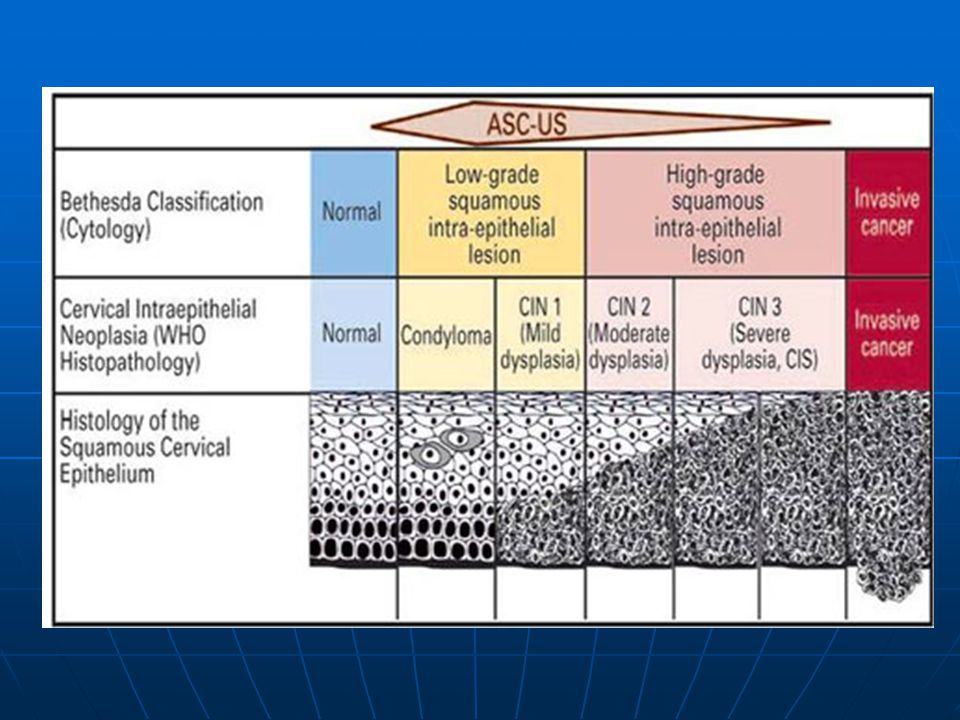
Τραχηλική ενδοεπιθηλιακή νεοπλασία (cervical intraepithelial neoplasia, CIN)
CIN I (ελαφρά δυσπλασία, LSIL):τα άτυπα κύτταρα περιορίζονται στο κατώτερο τριτημόριο του επιθηλίου, ενώ τα ανώτερα 2/3 παρουσιάζουν φυσιολογική διαφοροποίηση και ωρίμανση με επιπέδωση των κυττάρων CIN II (μέτρια δυσπλασία, HSIL): τα άτυπα κύτταρα καταλαμβάνουν το κάτω μισό του επιθηλίου,ενώ στο άνω μισό διακρίνονται στοιχεία διαφοροποίησης και ωρίμανσης. Οι πυρηνικές ανωμαλίες μπορεί να εκτείνονται σε όλο το πάχος του επιθηλίου είναι όμως εντονότερες στο κατώτερο μισό όπου μπορεί να εμφανισθούν αυξημένες μιτώσεις, εν μέρει άτυπες CIN III (έντονη δυσπλασία, HSIL, καρκίνωμα in situ):τα άτυπα κύτταρα καταλαμβάνουν όλο το πάχος του επιθηλίου χωρίς να παρουσιάζουν ουσιώδη διαφοροποίηση και ωρίμανση στην επιφάνεια. Πυρηνοκινησίες, άτυπες ή μη, εμφανίζονται σε όλες τις στιβάδες του επιθηλίου, οι δε επιθηλιακές αλλοιώσεις μπορεί να επεκτείνονται κατά μήκος των κρυπτών του ενδοτραχήλου
:τα άτυπα κύτταρα περιορίζονται στο κατώτερο τριτημόριο του επιθηλίου, ενώ τα ανώτερα 2/3 παρουσιάζουν φυσιολογική διαφοροποίηση και ωρίμανση με επιπέδωση των κυττάρων. CIN II (μέτρια δυσπλασία, HSIL): τα άτυπα κύτταρα καταλαμβάνουν το κάτω μισό του επιθηλίου,ενώ στο άνω μισό διακρίνονται στοιχεία διαφοροποίησης και ωρίμανσης. Οι πυρηνικές ανωμαλίες μπορεί να εκτείνονται σε όλο το πάχος του επιθηλίου είναι όμως εντονότερες στο κατώτερο μισό όπου μπορεί να εμφανισθούν αυξημένες μιτώσεις, εν μέρει άτυπες. CIN III (έντονη δυσπλασία, HSIL, καρκίνωμα in situ):τα άτυπα κύτταρα καταλαμβάνουν όλο το πάχος του επιθηλίου χωρίς να παρουσιάζουν ουσιώδη διαφοροποίηση και ωρίμανση στην επιφάνεια. Πυρηνοκινησίες, άτυπες ή μη, εμφανίζονται σε όλες τις στιβάδες του επιθηλίου, οι δε επιθηλιακές αλλοιώσεις μπορεί να επεκτείνονται κατά μήκος των κρυπτών του ενδοτραχήλου.")
21
HPV και καρκίνος τραχήλου
Ο κύριος αιτιολογικός παράγοντας στην ανάπτυξη δυσπλασίας του τραχήλου. HPV DNA ανευρίσκεται στο 99% των διηθητικών καρκινωμάτων του τραχήλου και στο 75-95% των υψηλόβαθμων ενδοεπιθηλιακών αλλοιώσεων (CIN II or III) Η εμμένουσα λοίμωξη είναι ο παράγοντας κλειδί στην εξέλιξη του καρκίνου. HPV λοίμωξη Έχουν αναγνωρισθεί πάνω από 100 υπότυποι του ιού >40 τύποι προσβάλουν την γεννητική περιοχή/τράχηλο “Υψηλού κινδύνου” ή ογκογόνοι τύποι 13 υπότυποι στο HC 2, εκ των οποίων οι 16, 18, 31, 45 προκαλούν τους περισσότερους καρκίνους παγκοσμίως. Επιπλέον 5 υπότυποι είναι πιθανόν καρκινογόνοι. Οι περισσότεροι ιοί υψηλού κινδύνου προκαλούν χαμηλόβαθμες αλλοιώσεις Χαμηλού κινδύνου υπότυποι: Σχετίζονται με καλοήθεις επιθηλιακές υπερπλαστικές αλλοιώσεις, όχι με διηθητικό καρκίνο. Εξωφυτικά κονδυλώματα τύποι 6 & 11
 Η εμμένουσα λοίμωξη είναι ο παράγοντας κλειδί στην εξέλιξη του καρκίνου. HPV λοίμωξη. Έχουν αναγνωρισθεί πάνω από 100 υπότυποι του ιού. >40 τύποι προσβάλουν την γεννητική περιοχή/τράχηλο. Υψηλού κινδύνου ή ογκογόνοι τύποι. 13 υπότυποι στο HC 2, εκ των οποίων οι 16, 18, 31, 45 προκαλούν τους περισσότερους καρκίνους παγκοσμίως. Επιπλέον 5 υπότυποι είναι πιθανόν καρκινογόνοι. Οι περισσότεροι ιοί υψηλού κινδύνου προκαλούν χαμηλόβαθμες αλλοιώσεις. Χαμηλού κινδύνου υπότυποι: Σχετίζονται με καλοήθεις επιθηλιακές υπερπλαστικές αλλοιώσεις, όχι με διηθητικό καρκίνο. Εξωφυτικά κονδυλώματα τύποι 6 & 11.")
22
HPV-λοίμωξη Μικροτραυματισμοί, μικροαμυχές του επιθηλίου→έκθεση των βασικών κυττάρων και διευκόλυνση της εισόδου του ιού 1-10 ιικά τεμαχίδια (virions)εισάγονται στο βασικό κύτταρο μέσω της α6-ιντεγκρίνης→μετακίνηση του ιικού γονιδιώματος στον πυρήνα του κυττάρου Στον πυρήνα τα ιικά γονιδιώματα είναι επισωματικά ξέχωρα από το DNA του κυττάρου ξενιστή Τα γονιδιώματα αντιγράφονται παράλληλα με το DNA του ξενιστή, διατηρώντας ένα χαμηλό αριθμό αντιγράφων (περίπου 50/ανά κύτταρο) και φτιάχνουν μια δεξαμενή προσβεβλημένων κυττάρων Το ιικό γονιδίωμα αντιγράφεται χρησιμοποιώντας δύο δικές του πρωτεΐνες (Ε1 και Ε2) και τη μηχανή μεταγραφής του DNA του ξενιστή Μετά την κυτταρική διαίρεση τα προσβεβλημένα θυγατρικά κύτταρα μεταναστεύουν προς την επιφάνεια όπου διαφοροποιούνται. Σε αντίθεση όμως με τα φυσιολογικά κερατινοκύτταρα τα οποία αποπίπτουν, αυτά εισέρχονται στη φάση S→πολλαπλασιασμός του ιικού γονιδιώματος σε χιλιάδες αντίγραφα ανά κύτταρο Έκφραση των γονιδίων L1 και L2 και σχηματισμός δομικών πρωτεϊνών (ενδιάμεση ζώνη) Το ιικό DNA πακετάρεται σε καψίδια που αποτελούνται από τις L1 και L2 πρωτεΐνες→ αθροίζονται στην επιφανειακή ζώνη→απελευθερώνονται στο εξωκυττάριο περιβάλλον με την αποφολίδωση των επιφανειακών κυττάρων→μόλυνση γειτονικών βασικών κυττάρων
εισάγονται στο βασικό κύτταρο μέσω της α6-ιντεγκρίνης→μετακίνηση του ιικού γονιδιώματος στον πυρήνα του κυττάρου. Στον πυρήνα τα ιικά γονιδιώματα είναι επισωματικά ξέχωρα από το DNA του κυττάρου ξενιστή. Τα γονιδιώματα αντιγράφονται παράλληλα με το DNA του ξενιστή, διατηρώντας ένα χαμηλό αριθμό αντιγράφων (περίπου 50/ανά κύτταρο) και φτιάχνουν μια δεξαμενή προσβεβλημένων κυττάρων. Το ιικό γονιδίωμα αντιγράφεται χρησιμοποιώντας δύο δικές του πρωτεΐνες (Ε1 και Ε2) και τη μηχανή μεταγραφής του DNA του ξενιστή. Μετά την κυτταρική διαίρεση τα προσβεβλημένα θυγατρικά κύτταρα μεταναστεύουν προς την επιφάνεια όπου διαφοροποιούνται. Σε αντίθεση όμως με τα φυσιολογικά κερατινοκύτταρα τα οποία αποπίπτουν, αυτά εισέρχονται στη φάση S→πολλαπλασιασμός του ιικού γονιδιώματος σε χιλιάδες αντίγραφα ανά κύτταρο. Έκφραση των γονιδίων L1 και L2 και σχηματισμός δομικών πρωτεϊνών (ενδιάμεση ζώνη) Το ιικό DNA πακετάρεται σε καψίδια που αποτελούνται από τις L1 και L2 πρωτεΐνες→ αθροίζονται στην επιφανειακή ζώνη→απελευθερώνονται στο εξωκυττάριο περιβάλλον με την αποφολίδωση των επιφανειακών κυττάρων→μόλυνση γειτονικών βασικών κυττάρων.")
23
Φυσική ιστορία της καρκινογένεσης στον τράχηλο
Εμμένουσα λοίμωξη HPV λοίμωξη τραχήλου εξέλιξη Προκαρκινω ματώδεις αλλοιώσεις διήθηση Λοίμωξη Φυσιολ. τράχηλος Καρκίνος Κάθαρση υποστροφή Ήπια κυτταρική ατυπία Σοβαρή κυτταρική ατυπία From Mark Schiffman, NCI

25
Φυσική πορεία της CIN Εμμονή Εξέλιξη σε CIS Εξέλιξη σε Ca 57% 32% 11%
Υποστροφή Εμμονή Εξέλιξη σε CIS Εξέλιξη σε Ca CIN 1 57% 32% 11% 1% CIN 2 43% 35% 22% 5% CIN 3 ~56% - ~12%

26
(flat condyloma): abnormal mitotic figure (green circle), numerous binucleated cells (yellow circle), koilocytotic cells (stars), superficial parakeratosis (red arrow).
: abnormal mitotic figure (green circle), numerous binucleated cells (yellow circle), koilocytotic cells (stars), superficial parakeratosis (red arrow).")
27
(flat condyloma): flat condyloma (A) and normal ectocervical epithelium (B) next to each other.
: flat condyloma (A) and normal ectocervical epithelium (B) next to each other.")
29
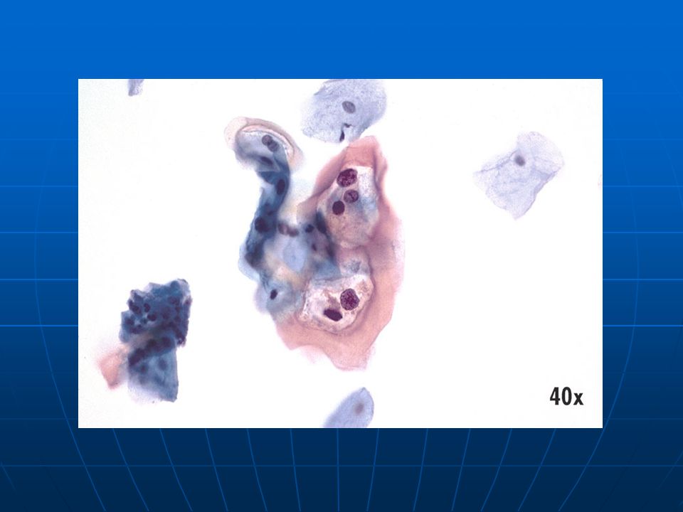
ASC-US • Atypical parakeratosis with pleomorphic nuclei and irregular cell shapes.

30
CIN 2 with koilocytosis: disorganized architecture of the lower half of the epithelium with cellular criteria of malignancy and viral infection

31
CIN 2 with koilocytosis: disorganized architecture of the lower half of the epithelium with cellular criteria of malignancy and viral infection

32
CIN 2 in situ hybridization, HPV 31 and 33 probes
CIN 2 in situ hybridization, HPV 31 and 33 probes. A few positive superficial cell nuclei.

33
HSIL – Moderate • Dense cytoplasm, sharp nuclear irregularities with ridges, indentations in nuclear membrane and coarse uniform chromatin.

34
CIN 3 Cervical intraepithelial neoplasia grade 3 (Richart) is a preneoplastic lesion of squamous epithelium corresponding to previous severe dysplasia and squamous intraepithelial carcinoma. Histologically, the proliferation of atypical parabasal cells, oriented in a vertical fashion, involves more than two-thirds. Architectural disorganization, typical or atypical mitotic figures, cytonuclear criteria of malignancy are present in more than the lower two thirds. Keratinization may be present, at the surface or in isolated cells inside the epithelium. Koilocytosis may be observed. CIN 3 superficial parakeratosis

35
CIN 2-3 The cellular organization of the epithelium is disturbed in the lower two-thirds and the cells display a high degree of nuclear and cellular abnormalities with typical and atypical mitoses.

36
CIN 3 the cellular organization of the epithelium is disturbed in more than the lower two-thirds and the cells display a high degree of nuclear and cellular abnormalities with typical and atypical mitoses.

37
CIN 3 superficial parakeratosis
CIN 3 superficial parakeratosis. Contrast with the normal exocervical epithelium

38
carcinoma in situ): endocervical glandular involvement.
: endocervical glandular involvement.")
39
HSIL – Severe • Small sheet of immature squamous cells with scant rim of cytoplasm, hyperchromatic nuclei with coarse, but evenly distributed chromatin as compared to normal population.

40
Τύποι καρκινωμάτων τραχήλου
Πλακώδες (ακανθοκυτταρικό) καρκίνωμα (70-75%) Αδενοκαρκίνωμα (15-25%) Αδενοπλακώδες (5-10%) Άλλοι σπάνιοι τύποι
 καρκίνωμα (70-75%) Αδενοκαρκίνωμα (15-25%) Αδενοπλακώδες (5-10%) Άλλοι σπάνιοι τύποι.")
41
Πλακώδες καρκίνωμα 3-5% των περιπτώσεων καρκίνου στις γυναίκες
Το 90% των πλακωδών καρκινωμάτων του τραχήλου ξεκινούν σε μια περιοχή 1 εκ. της ζώνης μετάπλασης Αναπτύσσεται γενικά σε υπόστρωμα CIN Μέση ηλικία εμφάνισης 50 έτη HPV 16 Πρωταρχικό σύμπτωμα→κολπική αιμορραγία Παραμελημένοι όγκοι→απόφραξη της ουροφόρου οδού λόγω διήθησης της ουροδόχου κύστης Μακροσκοπικά: πρώιμες αλλοιώσεις→ανώμαλες περιοχές του τραχηλικού βλεννογόνου προχωρημένες βλάβες→ανθοκραμβοειδή εξελκωμένα μορφώματα που καταστρέφουν τον τράχηλο Χαρακτηρίζονται ανάλογα με τον κυτταρικό τύπο που επικρατεί Μη κερατινοποιημένο από μεγάλα κύτταρα (η πλειονότητα) Κερατινοποιημένο από μεγάλα κύτταρα Πλακώδες καρκίνωμα από μικρά κύτταρα (χειρότερη πρόγνωση)
 Κερατινοποιημένο από μεγάλα κύτταρα. Πλακώδες καρκίνωμα από μικρά κύτταρα (χειρότερη πρόγνωση)")
42
Παράγοντες κινδύνου Συνουσία: χαμηλή επίπτωση στις παρθένες
Ηλικία πρώτης συνουσίας: πρώτη επαφή πριν τα 17 έτη Σεξουαλικά μεταδιδόμενα νοσήματα Κοινωνικοοικονομική κατάσταση: κατώτερες κοινωνικές ομάδες Κάπνισμα: καταστολή της τοπικής ανοσοεπαγρύπνισης των κυττάρων Langerhans HPV 16,18,33,….αδρανοποίηση προϊόντων ογκοκατασταλτικών γονιδίων HIV

43
Πρόγνωση πλακώδους καρκινώματος
Στάδιο 5ετής επιβίωση Βαθμός τοπικής διήθησης Ι 90% Περιοριζόμενο στον τράχηλο ΙΙ 75% Διήθηση του ανώτερου τμήματος του κόλπου ή των παρακείμενων παραμήτριων ιστών ΙΙΙ 30% Διασπορά στο πλευρικό τοίχωμα της πυέλου, τον κατώτερο κόλπο ή τους ουρητήρες IV 10% Επέκταση στο ορθό, το τοίχωμα της ουροδόχου ή εξωπυελικά

44
Μικροδιηθητικό πλακώδες καρκίνωμα (στάδιο ΙΑ)
Διήθηση σε βάθος <3 χιλ. κάτω από τη βασική μεμβράνη Απουσία αγγειακής διήθησης Απουσία λεμφαδενικών μεταστάσεων Απλή υστερεκτομή

45
Early stromal invasion Invasion of the connective tissue by a few mature neoplastic cells (less than 1 mm), invasion surrounded by a dense inflammatory infiltrate. Semi serial sections are needed. Histological entity integrated into microinvasive carcinomas since 2003 WHO classification. conization, presence of an early stromal invasion at the base of a gland (mature neoplastic squamous cells). Invasion demarcated by a lymphocytic infiltrate (circle).
. Invasion demarcated by a lymphocytic infiltrate (circle)..")
46
ESI conization, invasion demarcated by a lymphocytic infiltrate

47
ESI demarcated by a lymphocytic infiltrate.

48
Microinvasive carcinoma: mature neoplastic squamous cell nests infiltrating the connective tissue 2mm in depth (bar).
.")
49
This is the gross appearance of a cervical squamous cell carcinoma that is still limited to the cervix (stage I). The tumor is a fungating red to tan to yellow mass.

50
Here is another cervical squamous cell carcinoma
Here is another cervical squamous cell carcinoma. Note the IUD string protruding from the cervix. This implies that someone could have done a Pap smear when it was inserted. There is a natural history of progression of dysplasia to carcinoma, so don't leave dysplasias alone.

51
This is a larger cervical squamous cell carcinoma which spread to the vagina. A total abdominal hysterectomy with bilateral salpingo-oopherectomy (TAH-BSO) was performed.
 was performed..")
52
This is why you do Pap smears--to prevent invasive squamous cell carcinomas from occurring. With Pap smears, pre-neoplastic and neoplastic cervical lesions can be detected when small and treated. Nests of squamous cell carcinoma have invaded underlying stroma at the center and left.

53
At high magnification, nests of neoplastic squamous cells are invaded through a chronically inflamed stroma. This cancer is well- differentiated, as evidenced by keratin pearls. However, most cervical squamous carcinomas are non-keratinizing.

54
ΜΗ ΚΕΡΑΤΙΝΟΠΟΙΗΜΕΝΟ

55
ΜΗ ΚΕΡΑΤΙΝΟΠΟΙΗΜΕΝΟ

56
ΜΗΚΕΡΑΤΙΝΟΠΟΙΗΜΕΝΟ

57
Poorly-differentiated invasive squamous cell carcinoma

58
Squamous Cell Carcinoma
• Single malignant cells. Dense polygonal cytoplasm. Nucleus very open, with parachromatin clearing, prominent nucleoli, irregularities in nuclear membrane. Incorporate all features into interpretation. Keratinized squamous cell carcinoma more straightforward; keratin debris may be identified.

59
Squamous Cell Carcinoma
• Dense, angular cytoplasm helps to identify this group of malignant cells as squamous in differentiation. Prominent nucleoli present, with irregular chromatin distribution and thickened nuclear membranes.

60
Αδενοκαρκίνωμα Προέρχεται από τα αδενικά κύτταρα του ενδοτραχήλου
Μπορεί να προηγείται αδενοκαρκίνωμα in situ HPV 18 Ηλικία εμφάνισης 56 έτη Δύσκολα ορατό κλινικά λόγω εντόπισης Έχει την τάση να δίνει νωρίτερα λεμφαδενικές μεταστάσεις και είναι λιγότερο ακτινοευαίσθητο

61
Glandular dysplasia A glandular lesion characterized by significant nuclear abnormalities that are more striking than those encountered in glandular atypia, but do not fulfill the criteria for adenocarcinoma in situ (WHO 2003). (See also: AGC) GD pseudo-stratification of the endocervical epithelial nuclei associated with striking nuclear atypia. Persistence of essentially normal glandular epithelium on a small portion of the gland. (arrow)
 GD pseudo-stratification of the endocervical epithelial nuclei associated with striking nuclear atypia. Persistence of essentially normal glandular epithelium on a small portion of the gland. (arrow).")
62
Pseudo-stratification of the endocervical epithelial nuclei associated with nuclear atypia.

63
Moderate endocervical dysplasia: sharp transition between normal and dysplastic glandular epithelium

64
Adenocarcinoma in situ: sharp transition between normal (+) and neoplastic endocervical epithelium (arrow
 and neoplastic endocervical epithelium (arrow")
65
in situ (left) coexisting with normal endocervical epithelium (right).
 coexisting with normal endocervical epithelium (right).")
66
in situ: contrast between a normal gland (left) and a neoplastic gland (right).
 and a neoplastic gland (right).")
67
AIS • This group of glandular cells exhibits the nuclear crowding as well as hyperchromatic chromatin pattern. Cell dyshesion is also evident along the edges.

68
Coexisting adenocarcinoma in situ (arrow) and CIN 3 (star).
 and CIN 3 (star).")
69
AIS of the uterine cervix (blue dashes) associated with CIN 3 (red dashes).
 associated with CIN 3 (red dashes).")
70
microinvasive adenocarcinoma with neoplastic glands seen outside the normal glandular field (for the staging see the TNM and FIGO classifications).
.")
71
Well-differentiated invasive endocervical adenocarcinoma (grade I) composed of tubules (with multi-layered epithelium) which infiltrate the connective tissue.
 composed of tubules (with multi-layered epithelium) which infiltrate the connective tissue.")
72
Poorly differentiated invasive endocervical adenocarcinoma (grade III).
.")
73
Endocervical Adenocarcinoma
• Cluster of glandular cells with scalloping, poly-engulfment and nucleoli.

74
Carcinoma with malignant glandular (arrow) and squamous components (star).
 and squamous components (star).")
75
Carcinoma with malignant glandular (arrow) and squamous components (star).
 and squamous components (star).")
76
Colposcopy of a normal cervix (Panel A), cervix with acetowhitening with arrow denoting acetowhite area (Panel B), and a cervix with invasive carcinoma (Panel C).
, cervix with acetowhitening with arrow denoting acetowhite area (Panel B), and a cervix with invasive carcinoma (Panel C).")
77
Γυναίκες κάτω των 30 Test Pap αρνητικό ASC-US HPV (-) HPV (+)
Επανάληψη 1 έτος κολποσκόπηση HPV (-) HPV (+) Επανάληψη 1 έτος κολποσκόπηση
 HPV (+) Επανάληψη. 1 έτος. κολποσκόπηση.")
78
Γυναίκες άνω των 30 HPV (-) Test pap αρνητικό HPV (-) Test pap ASC-US
επανάληψη 3 έτη επανάληψη 1 έτος κολποσκόπηση

79
Επανάληψη και των 2 σε 6-12 μήνες
Γυναίκες άνω των 30 HPV+ Test pap αρνητικό HPV+ Test pap ASC-US HPV+ Test pap αλλοίωση κολποσκόπηση κολποσκόπηση Επανάληψη και των 2 σε 6-12 μήνες Αμφότερα αρνητικά: επανέλεγχος σε 3 χρόνια Κυτταρολογική ASC-US/HPV (-): επανάληψη σε 12 μήνες Κυτταρολογική με αλλοιώσεις/ HPV – ή +: κολποσκόπηση HPV+/οποιοδήποτε αποτέλεσμα κυτταρολογικής: κολποσκόπηση
: επανάληψη σε 12 μήνες. Κυτταρολογική με αλλοιώσεις/ HPV – ή +: κολποσκόπηση. HPV+/οποιοδήποτε αποτέλεσμα κυτταρολογικής: κολποσκόπηση.")
Παρόμοιες παρουσιάσεις
, είναι δύο μέλη της οικογένειας του ιού του έρπητα, herpesviridae, που μολύνει.>")


 Έχουν τη δική τους ιδιαίτερη οσμή. Με την παρουσία οξειδίων του αζώτου και ηλιακού.>")

