Κατέβασμα παρουσίασης
Η παρουσίαση φορτώνεται. Παρακαλείστε να περιμένετε

1
Μικροβιακές οφθαλμικές λοιμώξεις
Επιπεφυκίτιδα Haemophilus influenzae Διάφορα άλλα μικρόβια Λόγω χρήσης μη αποστειρωμένων φακών επαφής Γονοκοκκική οφθαλμία στα νεογέννητα Neisseria gonorrhoeae Μεταδίδεται κατά τη δίοδο του νεογεννήτου απ΄πο το γεννητικό σωλήνα

2
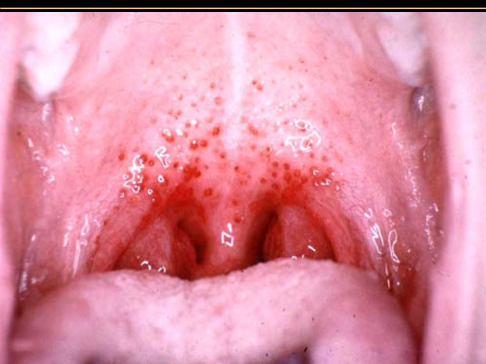
Μικροβιακές οφθαλμικές λοιμώξεις
Chlamydia trachomatis Επιπεφυκίτιδα Μεταδίδεται κατά τη γέννηση Μετάδοση από νερά κολυμβητικών δεξαμενών Αντιβιοτικό εκλογής: Τετρακυκλίνες Τράχωμα (λοίμωξη κερατοειδούς) Υψηλότερη αιτία τύφλωσης παγκοσμίως Μόνιμες βλάβες στον κερατοειδή
 Υψηλότερη αιτία τύφλωσης παγκοσμίως. Μόνιμες βλάβες στον κερατοειδή.")
3
Μικροβιακές οφθαλμικές λοιμώξεις
Figure 21.20

4
Μικροβιακές οφθαλμικές λοιμώξεις
Ερπητική κερατίτιδα Herpes simplex virus 1 (HHV-1) Προσβολή κερατοειδούς, μπορεί να οδηγήσει σε τύφλωση Θεραπεία με τριφλουριδίνη Acanthamoeba κερατίτιδα Μεταδίδεται από το νερό Φακοί επαφής
 Προσβολή κερατοειδούς, μπορεί να οδηγήσει σε τύφλωση. Θεραπεία με τριφλουριδίνη. Acanthamoeba κερατίτιδα. Μεταδίδεται από το νερό. Φακοί επαφής.")
5
Μικροβιακές οφθαλμικές λοιμώξεις
Figure 21.21

6
Λοιμώξεις ανώτερου αναπνευστικού
Λαρυγγίτιδα-Φαρυγγίτιδα: S. pneumoniae, S. pyogenes, ιοί Αμυγδαλίτιδα: S. pneumoniae, S. pyogenes, ιοί Ιγμορίτιδες: Βακτήρια Επιγλωττίτιδα: H. influenzae

7
Λοιμώξεις ανώτερου αναπνευστικού
Από δυνητικά παθογόνα Figure 24.1

8
Streptococcal pharyngitis (Strep throat)
Streptococcus pyogenes Resistant to phagocytosis Streptokinases lyse clots Streptolysins are cytotoxic Diagnosis by indirect agglutination Figure 24.3

10
Group A Beta Hemolytic Streptococcus

11
BETA HEMOLYSIS

12
Επιπλοκές φαργγίτιδας από Στρεπτόκοκκο Ομάδας Α
Μέση ωτίτιδα Ιγμορίτιδες Περιαμυγδαλιδικό ή φαρυγγικό απόστημα Τραχηλική αδενίτιδαs

13
Streptococcal Cervical Adenitis

14
Scarlet Fever (Οστρακιά)
Streptococcus pyogenes Pharyngitis Erythrogenic toxin produced by lysogenized S. pyogenes Figure 24.4

15
DISEASES Impetigo Erysipelas

16
DISEASES cont. Erysipelas Tonsillitis

17
DISEASES cont. Scarlet Fever Toxic Shock

18
Diphtheria Corynebacterium diphtheriae: Gram-positive rod
Diphtheria membrane of fibrin, dead tissue, and bacteria Diphtheria toxin produced by lysogenized C. diphtheriae Prevented by DTaP and Td vaccine (Diphtheria toxoid) Cutaneous diphtheria: Infected skin wound leads to slow healing ulcer
 Cutaneous diphtheria: Infected skin wound leads to slow healing ulcer.")
19
I. Organism -G+, club shaped, pleomorphic, aerobic rod

20
I. Organism -G+, club shaped, pleomorphic, aerobic rod
-metachromatic polyphosphate granules

21
I. Organism -G+, club shaped, pleomorphic, aerobic rod
-metachromatic polyphosphate granules -reduce potassium tellurite to tellurium metal - black ppt. on tellurite blood agar

22
I. Organism -G+, club shaped, pleomorphic, aerobic rod
-metachromatic polyphosphate granules -reduce potassium tellurite to tellurium metal - black ppt. on tellurite blood agar -subtypes gravis, intermedius, mitis: severity of infection differs depending on growth rate of subtype

23
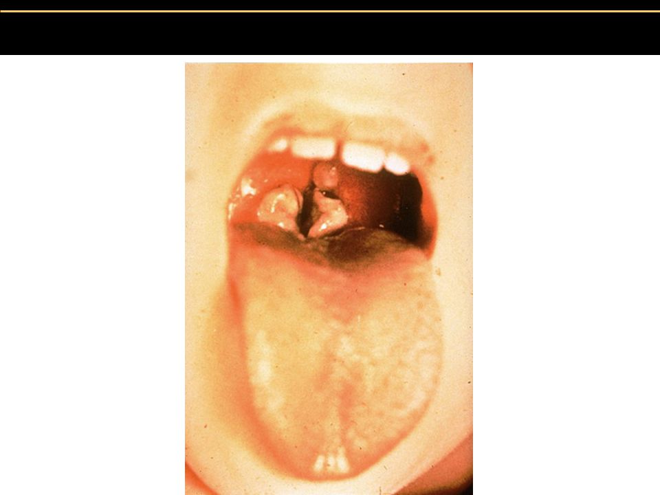
II. Clinical -usually presents as throat infection w/ sore throat, fever, sometimes swollen lymph glands -”pseudomembrane” forms at back of throat - may obstruct airway (“la garottilla”)
")
25
Pseudomembrane in Diptheria

27
Anatomy of the Ear

28
Anatomy of the Ear 3 Semicircular canals 4 Cochlea
1 Tympanic Membrane 2 Maleus Incus Stapes 3 Semicircular canals 4 Cochlea 5 Cochlear Nerve 6 Oval Window 7 Eustachian Tube 8 Orifice 9 Round Window10 Middle Ear Cavity

29
Otitis externa Otitis media Ear infections
tympanic membrane Otitis externa Otitis media Exogenous organisms via external auditory canal Endogenous organisms via eustachian tube

30
Otitis Media Epidemiology and Pathophysiology:
age - almost all children have one or more episodes before age 6 about 10% of children develop OM by age 3 months peak incidence between ages 6 and 15 months

31
Otitis Media Etiology: abnormal function of the eustachian tube

32
Otitis Media Microbiology (AOM) Streptococcus pneumoniae 35%
Haemophilus Influenzae % Moraxella catarrhalis % Alpha-hemolytic streptlococci % GAB-hemolytic streptococci % Staphylococcus aureus % Psuedomonas aeruginosa % Treated with broad-spectrum antibiotics Incidence of S. pneumoniae reduced by vaccine

33
Otits Media Microbiology (COM) Haemophilus influenzae 15%
Moraxella catarrhalis % Streptococcus pneumoniae % Alpha-hemolytic streptococci 3% Staphylococcus aureus % Pseudomonas artuginosa 2% GAB hemolytic streptococci %

34
Otitis Media Inflamation of the middle ear
Acute Otitis Media (AOM) - rapid onset of redness and bulging of the tympanic membrane, decreased mobility, pain, perforation with otorrhea
 - rapid onset of redness and bulging of the tympanic membrane, decreased mobility, pain, perforation with otorrhea.")
35
Otitis Media Figure 25.7

36
AOE: Mild to Moderate Stage
Progressive infection Symptoms Pain Increased pruritus Signs Erythema Increasing edema Canal debris, discharge

37
AOE: Severe Stage Severe pain, worse with ear movement Signs
Lumen obliteration Purulent otorrhea Involvement of periauricular soft tissue

38
Otitis Externa

39
COE: Signs Asteatosis Dry, flaky skin Hypertrophied skin
Mucopurulent otorrhea (occasional)
")
40
Otomycosis: Signs Canal erythema Mild edema
White, gray or black fungal debris

41
Otomycosis

42
Herpes Zoster Oticus: Symptoms
Early: burning pain in one ear, headache, malaise and fever Late (3 to 7 days): vesicles, facial paralysis
: vesicles, facial paralysis.")
43
Erysipelas Acute superficial cellulitis
Group A, beta hemolytic streptococci Skin: bright red; well-demarcated, advancing margin Rapid treatment with oral or IV antibiotics if insufficient response

44
Otitis externa Organisms - Pseudomonas aeruginosa
- Staphylococcus aureus - Aspergillus spp. Treatment - oral antibiotics – NO! - topical broad spectrum (thick drops or wicks) - cotricosteroids Prevention - avoid predisposing events. ( post swim drops, blue tack, ear “ NO GO” zone)
 - cotricosteroids. Prevention - avoid predisposing events. ( post swim drops, blue tack, ear NO GO zone)")
45
Sinusitis 32 million cases annually in U.S.
% of colds are complicated by sinusitis Most acute sinusitis occurs from viral and bacterial co-infection

46
Sinusitis

47
Sinusitis

48
Sinusitis Minor Symptoms Major symptoms Headache Facial Pain*
Nasal Obstruction Nasal Discharge Postnasal Drainage Hyposmia Fever Minor Symptoms Headache Halitosis Fatigue Oral Pain Cough Ear pain, pressure, fullness *Location = diagnosis

49
Sinusitis Microbiology
S. pneumoniae and H. influenzae cause 70% of sinusitis Anaerobes, S. aureus and M.catarrhalis cause most of remaining 30%

50
Common cold Rhinoviruses (50%) Coronaviruses (15-20%)
Rhinoviruses attached to ICAN-1 on nasal mucosa

51
Microbial Diseases of the Lower Respiratory System
Bacteria, viruses, & fungi cause: Bronchitis Bronchiolitis Pneumonia

52
Lower Respiratory System
The ciliary escalator keeps the lower respiratory system sterile. Figure 24.2

53
Pertussis (Whooping Cough)
Bordetella pertussis: Gram-negative coccobacillus Capsule Tracheal cytotoxin of cell wall damaged ciliated cells Pertussis toxin Prevented by DTaP vaccine (acellular Pertussis cell fragments) Figure 24.8
 Figure")
54
Pertussis (Whooping Cough)
Stage 1: Catarrhal stage, like common cold Stage 2: Paroxysmal stage: Violent coughing sieges Stage 3: Convalescence stage

55
Tuberculosis Mycobacterium tuberculosis: Acid-fast rod. Transmitted from human to human M. bovis: <1% U.S. cases, not transmitted from human to human M. avium-intracellulare complex infects people with late stage HIV infection Figure 24.9

56
Tuberculosis Figure

57
Tuberculosis Figure

58
Tuberculosis Figure

59
Tuberculosis Treatment of Tuberculosis: Prolonged treatment with multiple antibiotics Vaccines: BCG, live, avirulent M. bovis. Not widely used in U.S.

60
Tuberculosis Diagnosis: Tuberculin skin test screening
+ = current or previous infection Followed by X-ray or CT, acid-fast staining of sputum, culturing bacteria Figure 24.11

61
Pneumomoccal Pneumonia
Streptococcus pneumoniae: Gram-positive encapsulated diplococci Diagnosis by culturing bacteria Penicillin is drug of choice Figure 24.13

62
Pneumomoccal Pneumonia

63
Haemophilus influenzae Pneumonia
Gram-negative coccobacillus Alcoholism, poor nutrition, cancer, or diabetes are predisposing factors Second-generation cephalosporins

64
Mycoplasmal Pneumonia
Mycoplasma pneumoniae: pleomorphic, wall-less bacteria Also called primary atypical pneumonia and walking pneumonia Common in children and young adults Diagnosis by PCR or by IgM antibodies Figure 24.14

65
Mycoplasmal Pneumonia
Figure 11.19a, b

66
Legionellosis Legionella pneumophila: Gram-negative rod
L. pneumophila is found in water Transmitted by inhaling aerosols, not transmitted from human to human Diagnosis: culturing bacteria Treatment: Erythromycin

67
Psittacosis (Ornithosis)
Chlamydia psittaci: gram-negative intracellular bacterium Transmitted by elementary bodies from bird dropping to humans Reorganizes into reticulate body after being phagocytized Diagnosis: culturing bacteria in eggs or cell culture Treatment: Tetracycline

68
Chlamydial Pneumonia Chlamydia pneumoniae
Transmitted from human to human Diagnosis by FA test Treatment: Tetracycline

69
Mycoplasmal pneumonia
Mycoplasma pneumoniae: pleomorphic, wall-less bacteria Also called primary atypical pneumonia and walking pneumonia Common in children and young adults Diagnosis by PCR or by IgM antibodies

70
Q fever Figure 24.15

71
Viral Pneumonia Viral pneumonia as a complication of influenza, measles, chickenpox Viral etiology suspected if no cause determined Respiratory Syncytial Virus (RSV) Common in infants; 4500 deaths annually Causes cell fusion (syncytium) in cell culture Symptoms: coughing Diagnosis by serologic test for viruses and antibodies Treatment: Ribavirin
 Common in infants; 4500 deaths annually. Causes cell fusion (syncytium) in cell culture. Symptoms: coughing. Diagnosis by serologic test for viruses and antibodies. Treatment: Ribavirin.")
72
Influenza Chills, fever, headache, muscle aches (no intestinal symptoms) 1% mortality due to secondary bacterial infections Treatment: Amantadine Vaccine for high-risk individuals

73
Influenza Hemagglutinin (H) spikes used for attachment to host cells
Neuraminidase (N) spikes used to release virus from cell
 spikes used to release virus from cell.")
74
Influenza Figure 24.16

75
Influenza Antigenic shift Changes in H and N spikes
Probably due to genetic recombination between different strains infecting the same cell Antigenic drift Mutations in genes encoding H or N spikes May involve only 1 amino acid Allows virus to avoid mucosal IgA antibodies

76
Influenza serotypes A: causes most epidemics, H3N2, H1N1, H2N2
B: moderate, local outbreaks C: mild disease

77
Histoplasmosis Histoplasma capsulatum, dimorphic fungus (a) 37˚
Figure 24.17

78
Pneumocystis Pneumonia
Pneumocystis jiroveci (P. carinii) found in healthy human lungs Pneumonia occurs in newly infected infants & immunosuppressed individuals Treatment: Timethoprim-sulfamethoxazole Figure 24.22
 found in healthy human lungs. Pneumonia occurs in newly infected infants & immunosuppressed individuals. Treatment: Timethoprim-sulfamethoxazole. Figure")
79
Blastomycosis Blastomyces dermatitidis, dimorphic fungus Found in soil
Can cause extensive tissue destruction Treatment: amphotericin B

80
Opportunistic fungi involved in respiratory disease:
Aspergillus Rhizopus Mucor Mucor rouxii Figure 12.2b, 12.4

Παρόμοιες παρουσιάσεις


. ΣΥΓΚΟΠΤΙΚΗ ΚΡΙΣΗ Αιφνίδια και σύντομη απώλεια συνείδησης που προκαλείται από ανεπαρκή εγκεφαλική αιμάτωση λόγω μειωμένης.>")


