Κατέβασμα παρουσίασης
Η παρουσίαση φορτώνεται. Παρακαλείστε να περιμένετε

1
ΑΓΓΕΙΟΓΡΑΦΙΚΗ ΣΟΥΙΤΑ ΟΡΓΑΝΩΣΗ ΚΑΙ ΑΚΤΙΝΟΠΡΟΣΤΑΣΙΑ
Αχιλλέας Χατζηιωάννου ABR,EBIR Καθηγητής Ακτινολογίας ΕΚΠΑ.

2
DEPARTMENT LAYOUT Total 65 m2 not including the X ray control
Large lead lined doors to allow admission of bed with ancillary equipment Ceiling 3,5 -4 m with additional space above to allow mechanical access

3
MAIN FEATURES Fluoroscopy unit
Dirty utility space adjoining the examination space Scrub sink with hot and cold water Surveillance X ray control (10 m2) Storage space, apron rack and shelves for all equipment Surgical ceiling mounted lights Patient preparation space/room
 Storage space, apron rack and shelves for all equipment. Surgical ceiling mounted lights. Patient preparation space/room.")
4
Fluoroscopy history Popular Science: July 1939

5
Fluoroscopy unit Motion features
C or U arm which can rotate in axial and sagittal planes Angles of rotation must be displayed on the monitor Ability to vary source detector distance Combination of fluoroscopy arm and table movement should make imaging of the whole body possible Manual override and locking should be possible

6
Fluoroscopy unit Table features
Support at least 140 kg of weight Tilting capability both in craniocaudal and lateral direction Ability to move away from C-arm in case of emergency Additional ( foot and hand) controls to use by the radiographer
 controls to use by the radiographer.")
7
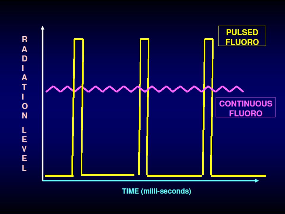
Fluoroscopy unit Imaging features
Image intensifier with large field of view (FOV) Digital subtraction and acquisition (DSA) Pulsed fluoroscopy for dose reduction Variety of frame rates Various collimators Filters to reduce skin dose Road mapping and land marking Last image hold and frame grab
 Digital subtraction and acquisition (DSA) Pulsed fluoroscopy for dose reduction. Variety of frame rates. Various collimators. Filters to reduce skin dose. Road mapping and land marking. Last image hold and frame grab.")
8
Fluoroscopy unit Image quality
The image quality mainly depends on the flat panel (image intensifier) and the processing system Image quality of fixed systems >> quality of mobile systems (difference in focal spot and tissue penetration)
 and the processing system. Image quality of fixed systems >> quality of mobile systems (difference in focal spot and tissue penetration)")
9
Fluoroscopy unit Image quality – Image Intensifier
The image intensifier determines the Field of View (FOV). FOV is of great importance especially in aortic interventions. It depends on the size of the image intensifier. The image intensifier should have: - large FOV ( cm) - three modes of magnification -spatial resolution at least 2.5 line pairs/mm in the 36-cm FOV, 3.3 line pairs/mm in the cm FOV, and 4.6 line pairs/mm in the 15-cm FOV - contrast ratio at least 20:1 - automatic brightness control
. FOV is of great importance especially in aortic interventions. It depends on the size of the image intensifier. The image intensifier should have: - large FOV ( cm) - three modes of magnification. -spatial resolution at least 2.5 line pairs/mm in the 36-cm FOV, 3.3 line pairs/mm in the 23- cm FOV, and 4.6 line pairs/mm in the 15-cm FOV. - contrast ratio at least 20:1. - automatic brightness control.")
10
Fluoroscopy unit Image quality – Processing system
Digital Subtraction and Acquisition 1024x1024 image matrix (ability to display compressed images on a 512x512 matrix) Display at least five frames per second in the 1024x1024 mode. Road-mapping (helpful during percutaneous transluminal angioplasty, with or without stent placement, and during passage of guidewires through tortuous vessels) Measurement of vessel diameter
 Display at least five frames per second in the 1024x1024. mode. Road-mapping (helpful during percutaneous transluminal angioplasty, with or without stent placement, and during passage of guidewires through tortuous vessels) Measurement of vessel diameter.")
11
Fluoroscopy unit Heat capacity
Overheating is rare in modern equipment due to the presence of excellent cooling systems (oil and water based)
")
12
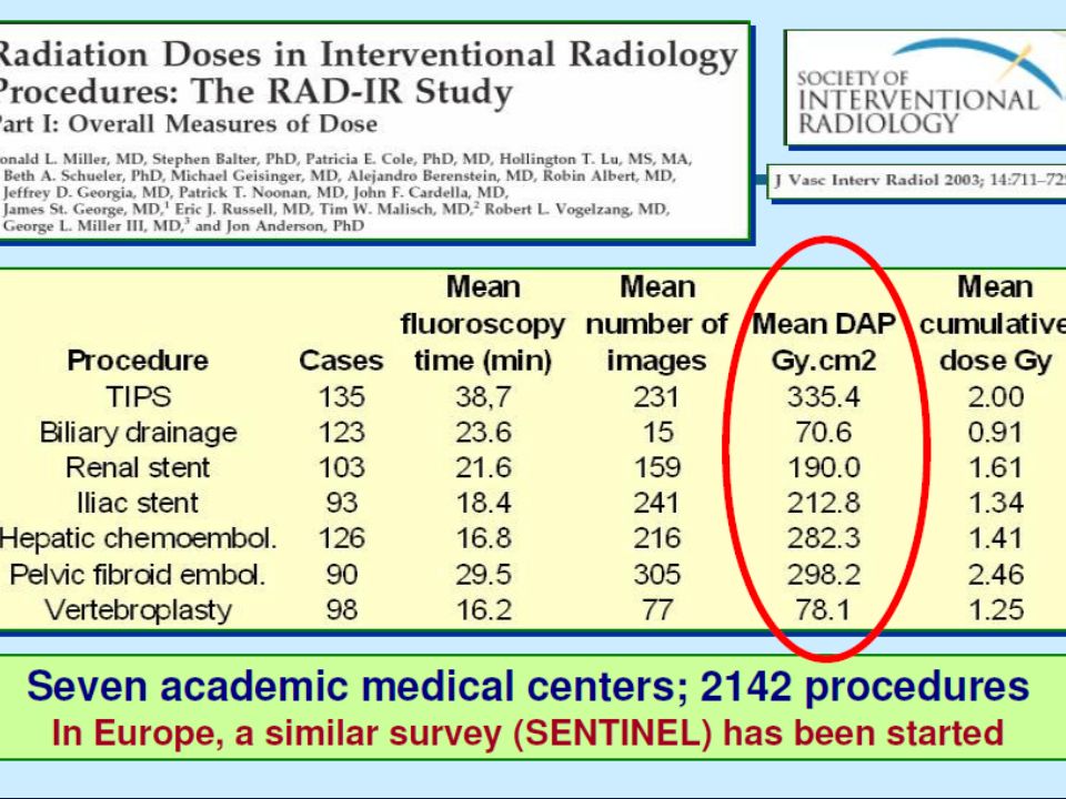
Fluoroscopy unit Radiation exposure
It varies greatly between different procedures It is dependent on multiple factors. One factor is the fluoroscopy unit itself. Radiation exposure is higher using portable C-arms than fixed systems

13
Don’t forget

14
Important issues of a modern angiosuite
Sterility issues Ability to convert Anaesthesia Ergonomics Personnel

15
Important issues in a modern angiosuite Sterility issues
Angiographic and interventional procedures should be performed in strictly aseptic conditions Primary and secondary operators should wear sterile gowns and gloves as well as surgical caps and masks. Sterile, impervious barriers should be available to cover any part of the equipment that may contaminate the field Appropriate receptacles for contaminated items should be readily available OR sterility > Angiosuite sterility

16
Important issues in a modern angiosuite Ability to convert
Majority of endovascular procedures without major intra-operative complications In case of conversion to open surgery, the lack of adequate lightning, suction, surgical and anaesthetic equipment and room for surgical instruments, and the extra personnel can make even small procedures challenging

17
Important issues in a modern angiosuite Anesthesia
Although endovascular procedures are minimally invasive,adequate monitoring of the cardiopulmonary system is essential in certain procedures (eg carotid stenting) The presence of an anesthesiologist, a specialized nurse and all anesthetic equipment seem to be of major importance
 The presence of an anesthesiologist, a specialized nurse and all anesthetic equipment seem to be of major importance.")
18
Important issues in a modern angiosuite Ergonomics
Poor ergonomics is one of the major drawbacks of mobile systems Ideally, a monitor should be placed just below eye level (10-15 degrees angle) Monitors on bothsides of the table Ceiling-fixed monitors perform a great range of positions,without claiming space on the floor. Adjustment of the position of these monitors is easier
 Monitors on bothsides of the table. Ceiling-fixed monitors perform a great range of positions,without claiming space on the floor. Adjustment of the position of these monitors is easier.")
19
Important issues in a modern angiosuite Personnel
Specially trained personnel Physicians should spend enough time doing procedures to fulfill the obligations of the angio suite and should perform a sufficient number of procedures to maintain proficiency and competence. All personnel should have knowledge about catheter techniques and the various materials that are used such as sheaths, guide wires, catheters and stent-grafts All personnel should have knowledge about radiation safety measures

20
ΧΕΡΙΑ ΑΚΤΙΝΟΛΟΓΟΥ

21
ΒΛΑΒΕΣ ΜΕΤΑ ΑΠΟ ΣΤΕΦΑΝΙΟΓΡΑΦΙΑ
[ 6 – 8 ΕΒΔΟΜΑΔΕΣ ΕΒΔΟΜΑΔΕΣ

22
ΔΙΑΦΟΡΕΣ ΒΛΑΒΕΣ ΜΕΤΑ FLUORO

24
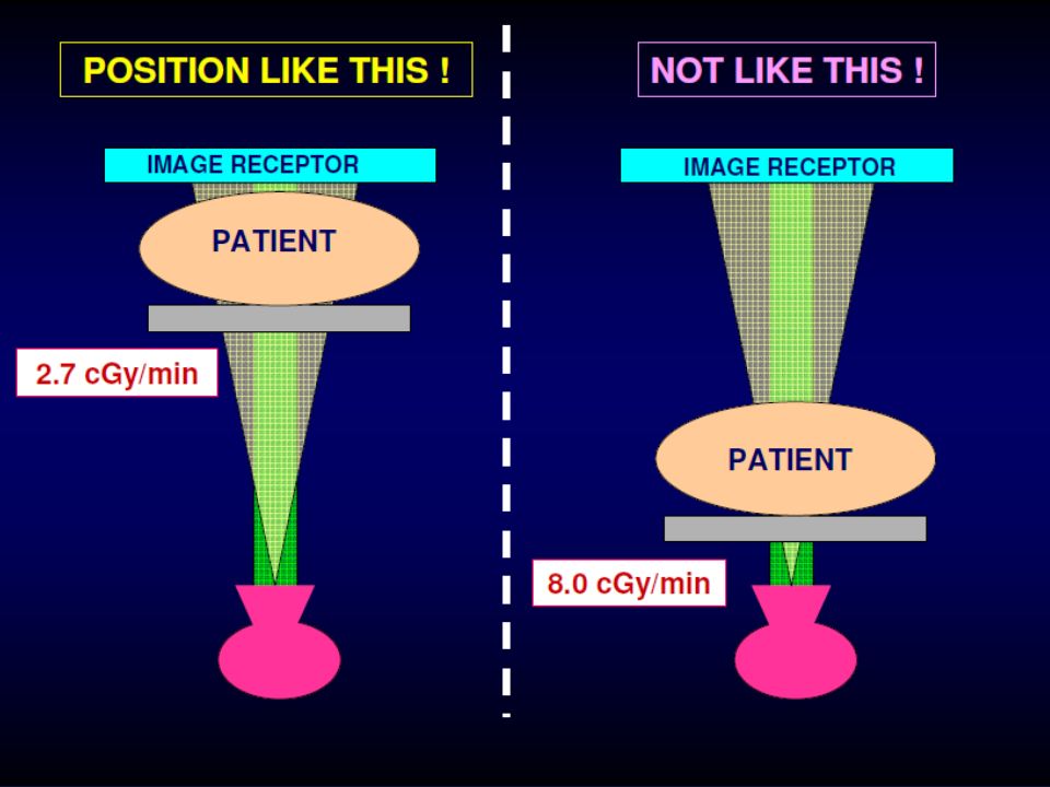
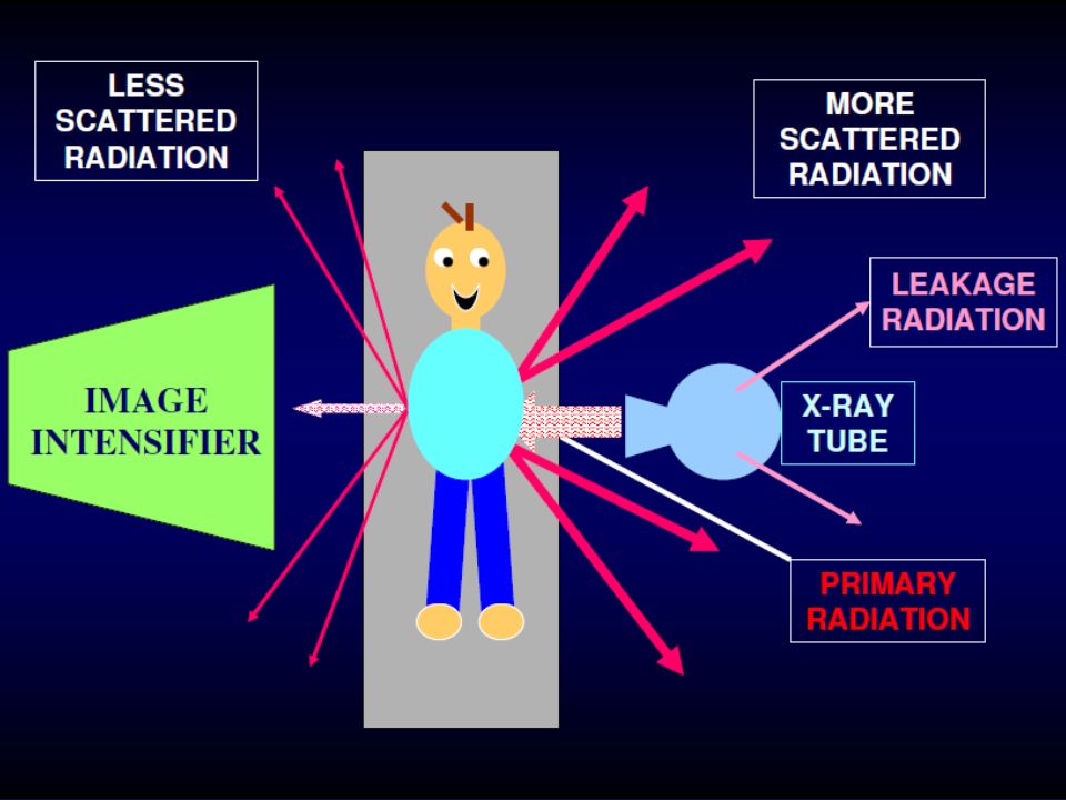
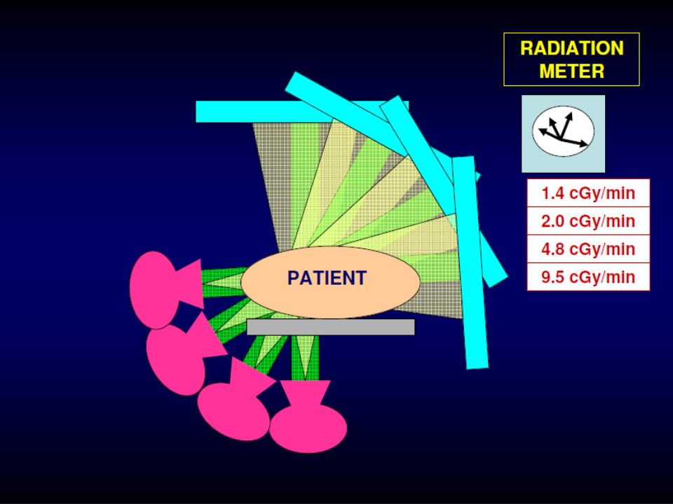
ΕΚΘΕΣΗ ΑΣΘΕΝΟΥΣ vs. ΕΠΕΜΒΑΤΙΣΤΗ
• Η απορροφούμενη ακτινοβολία από τον ασθενή και από τον επεμβατιστή είναι ανάλογες : – Μεγαλύτερη ακτινοβόληση του ασθενούς = μεγαλύτερη διάχυση ακτινοβολίας και ακτινοβόληση του επεμβατιστή • Σε απόσταση 1 μέτρου χωρίς θωράκιση, ο ρυθμός ακτινοβόλησης από σκεδαζόμενη ακτινοβολία είναι περίπου – φορές της ακτινοβολίας που λαμβάνει ο ασθενής στον ίδιο χρόνο • Συνεπώς μειώνοντας τον χρόνο και την ένταση ακτινοβόλησης του ασθενούς, μειώνεται και η δόση που λαμβάνει ο επεμβατιστής κατά την επέμβαση.

28
ΕΠΙΔΡΑΣΗ ΧΑΜΗΛΗΣ ΔΟΣΕΩΣ
• Deterministic effects – Dose dependent – Presumed to have a threshold dose • Stochastic effects (e.g. DNA damage) – Leukemia – Thyroid cancer
 – Leukemia. – Thyroid cancer.")
29
Deterministic Effects
• Cataracts – Observed at mSv ( REM) • Hair loss or skin erythema – Acute phase reaction at mSv – 65% of TACE cases exceed 2000mSv – Many procedures where skin dose at threshold • X-ray dose is cumulative so both these effects can occur in operators as well N Hidajat. CVIR 2006; 29:
 • Hair loss or skin erythema. – Acute phase reaction at mSv. – 65% of TACE cases exceed 2000mSv. – Many procedures where skin dose at threshold. • X-ray dose is cumulative so both these effects can occur in operators as well. N Hidajat. CVIR 2006; 29:")
30
Are The Dose Limits Low Enough?
• 59 interventional radiologists screened • Signs of radiation-related lens changes (posterior subcapsular cataracts) were found in five (8 percent) • Other authors have suggested threshold for early changes may be as low as 100 mSv Junk A, Haskal ZJ, Machan L, Worgul B. SIR Annual Meeting. Phoenix AZ
 were found in five (8 percent) • Other authors have suggested threshold for early changes may be as low as 100 mSv. Junk A, Haskal ZJ, Machan L, Worgul B. SIR Annual Meeting. Phoenix AZ.")
31
Radiation Induced Cataract
From: Vañó E, Gonzalez L, Beneytez F, Moreno F. Lens injuries induced by occupational exposure in non-optimized interventional radiology laboratories. BJR 1998; 71:

33
Dose To Lower Legs Causing Hair Loss?

35
ΠΕΡΙΟΡΙΣΜΟΣ ΧΡΟΝΟΥ ΑΚΤΙΝΟΣΚΟΠΗΣΗΣ & ΑΡΙΘΜΟΥ DSA ΕΙΚΟΝΩΝ
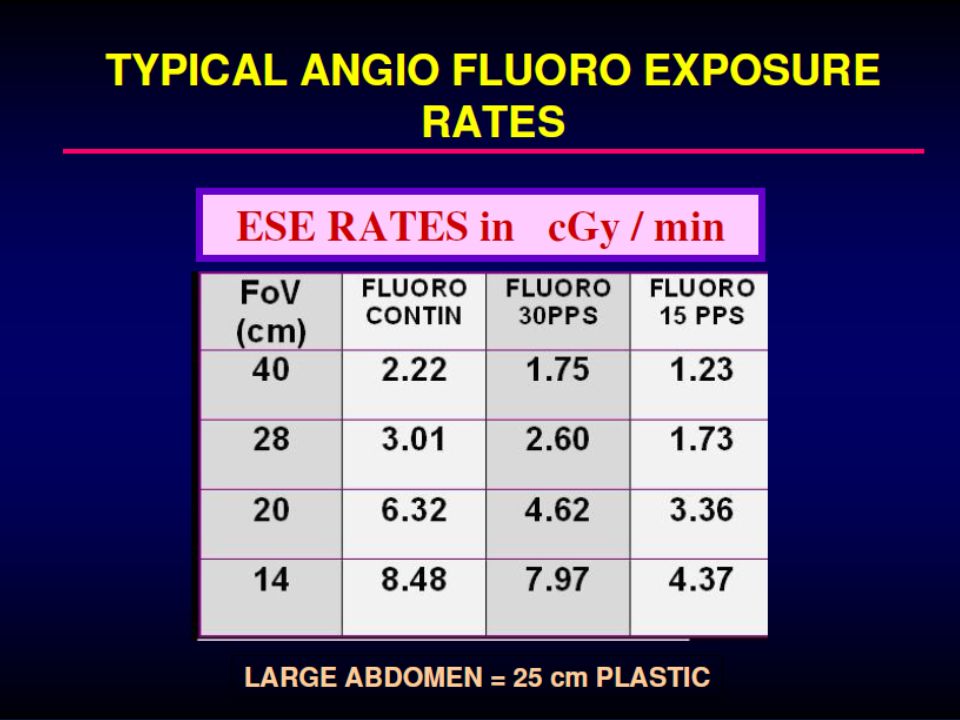
• 1 ΛΕΠΤΟ FLUORO ΙΣΟΔΥΝΑΜΕΙ ΜΕ 1 – 5 cGy ΣΤΟΝ ΑΣΘΕΝΗ – ΧΡΗΣΗ < 30 – 40 MIN FLUORO • ΧΡΗΣΗ ΤΕΛΕΥΤΑΙΑΣ ΕΙΚΟΝΑΣ ΓΙΑ ΜΕΛΕΤΗ ΤΗΣ ΑΝΑΤΟΜΙΑΣ – ΌΧΙ ΣΥΝΕΧΗΣ ΑΚΤΙΝΟΣΚΟΠΗΣΗ – ΠΡΟΣΧΕΔΙΑΣΜΟΣ ΕΠΕΜΒΑΣΗΣ

36
DSA ΣΥΜΜΕΤΕΧΕΙ ΣΤΗΝ ΔΟΣΗ ΤΟΥ ΑΣΘΕΝΟΥΣ

38
ΣΩΣΤΗ ΧΡΗΣΗ

40
Ακτινοσκόπηση & Δόση Ακτινοβολίας
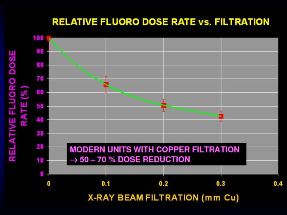
• Αποφυγή ακτινοσκόπησης υψηλής έντασης (high pulse rate) • Η αύξηση της μεγέθυνσης στην ακτινοσκόπηση αυξάνει το ποσό ακτινοβολίας • Η επιλογή χαμηλότερης έντασης δέσμης στην ακτινοσκόπηση μειώνει δραστικά την ακτινοβόληση του ασθενούς άρα και του επεμβατιστή • Το φιλτράρισμα στην ακτινοσκοπική λυχνία βελτιώνει την διαπερατότητα της ακτινοβολίας
 • Η αύξηση της μεγέθυνσης στην ακτινοσκόπηση αυξάνει το ποσό ακτινοβολίας. • Η επιλογή χαμηλότερης έντασης δέσμης στην ακτινοσκόπηση μειώνει δραστικά την ακτινοβόληση του ασθενούς άρα και του επεμβατιστή. • Το φιλτράρισμα στην ακτινοσκοπική λυχνία βελτιώνει την διαπερατότητα της ακτινοβολίας.")
46
Κοινά λάθη που κάνουν τα αποτελέσματα της επαγγελματικής έκθεσης σε ακτινοβολία αναξιόπιστα
• Ελλιπής χρήση των προσωπικών δοσιμέτρων. • Λάθη στην χρήση των δοσιμέτρων πάνω από και κάτω από την ποδιά (αλλαγή θέσης). • Χρήση των δοσιμέτρων και από άλλους επεμβατιστές (πχ όταν μένουν πάνω στην ποδιά).
. • Χρήση των δοσιμέτρων και από άλλους επεμβατιστές (πχ όταν μένουν πάνω στην ποδιά).")
48
ΚΑΙΝΟΤΟΜΙΑ ΕΙΝΑΙ ΤΟ ΜΕΛΛΟΝ ΜΑΣ
ΕΠΕΜΒΑΤΙΚΗ ΑΚΤΙΝΟΛΟΓΙΑ ΚΑΙΝΟΤΟΜΙΑ ΕΙΝΑΙ ΤΟ ΜΕΛΛΟΝ ΜΑΣ

Παρόμοιες παρουσιάσεις


 Διδάσκων: Καθηγητής Χρήστος.>")


 Όραση Μαρία Κουτρομάνου. Structure of the Eye: Iris The iris is similar to the diaphragm in a camera Your iris widens in dim light and.>")


