Κατέβασμα παρουσίασης
Η παρουσίαση φορτώνεται. Παρακαλείστε να περιμένετε

1
Ανατομία dsfsf dsfsf Αναπνευστικό Σύστημα dsfsf Ελευθερία Θωμαΐδου ,Pt

2
Respiratory tract Diameter (mm) 20-25 12-16 Conducting zone 1-12 0.5-1
Respiratory zone Diameter (mm) 20-25 12-16 1-12 0.5-1 < 0.5 0.3 diameter & cartilage smooth muscle Fig Stanfield
 < diameter & cartilage. smooth muscle. Fig Stanfield.")
3
Functions of the conducting zone
Provide a passageway for air to enter the respiratory zone Holds ‘dead space’ (~150 ml) Adjust air temperature Humidify air
 Adjust air temperature. Humidify air.")
4
Structure of the respiratory zone
Respiratory bronchioles Alveolar ducts Alveoli (alveolus) Alveolar sacs (in cluster)
 Alveolar sacs (in cluster)")
5
Structure of the respiratory zone
terminal bronchiole alveolar duct sacs respiratory bronchiole capillary network alveoli (a) (b) Fig Standfield
 (b) Fig Standfield.")
6
Function of the respiratory zone
Gas exchange between air and blood Location – respiratory membrane Mechanism – by simple diffusion

7
Structure of the thoracic cavity
lung intercostal muscle rib lung intercostal muscles pleural sac visceral pleura parietal pleura diaphragm intrapleural space pleural sac Chest wall (rib cage, sternum, thoracic vertebrae, connective tissue, intercostal muscles) Fig Stanfield
 Fig Stanfield.")
8
Pulmonary Pressures Pressure within the lungs is called intrapulmonary, or intra-alveolar, pressure. Between breaths = atmospheric pressure (760 mmHg) Inspiration = the volume of the thoracic cavity ↑ causing intrapulmonary pressure to ↓ below atmospheric pressure. This is also known as a negative pressure. Since air moves from areas of high to low air pressure, air flows into the lungs.
 Inspiration = the volume of the thoracic cavity ↑ causing intrapulmonary pressure to ↓ below atmospheric pressure. This is also known as a negative pressure. Since air moves from areas of high to low air pressure, air flows into the lungs.")
9
Pulmonary Pressures Intrapleural pressure – the pressure within the pleural cavity Intrapleural pressure – always negative, which acts like a suction to keep the lungs inflated Three main factors: 1. The surface tension of the alveolar fluid. This tends to pull each of the alveoli inward and therefore pulls the entire lung inward. Surfactant reduces this force.

10
Pulmonary Pressures 2. The elasticity of the lungs.
The abundant elastic tissue in the lungs tends to recoil and pull the lung inward. As the lung moves away from the thoracic wall, the cavity becomes slightly larger. 3. The elasticity of the thoracic wall. The elastic thoracic wall pulls away from the lung, further enlarging the pleural cavity and creating this negative pressure.

11
Pulmonary pressures at rest
Patm 760 mmHg (0 as ref) chest wall pleural sac Pip 756 mmHg (-4 rel) Palv 760 mmHg (0 rel) lung pleural sac Chest wall FRC diaphragm Palv = 0 mmHg Pip = - 4 mmHg Fig Germann
 chest wall. pleural sac. Pip 756 mmHg (-4 rel) Palv 760 mmHg (0 rel) lung. pleural. sac. Chest. wall. FRC. diaphragm. Palv = 0 mmHg. Pip = - 4 mmHg. Fig Germann.")
12
Inspiration and expiration
Inspiratory is active – muscles contract under nerve control Normal (quiet) expiration is passive Forceful expiration involves contraction of expiratory muscles (active expiration)
 expiration is passive. Forceful expiration involves contraction of expiratory muscles (active expiration)")
13
Muscles of ventilation
Inspiration Expiration Fig Stanfield

14
Mechanics of breathing
Movement of air occurs via bulk flow Movement of molecules due to pressure difference Inspiration Diaphragm pushes downward, ribs lift outward External intercostal muscles contract Volume of lungs increases Intrapulmonary pressure lowered Expiration Diaphragm relaxes, ribs pulled downward Volume of lungs decreases Intrapulmonary pressure raised

15
Changes in alveolar pressure & volume
Inspiration Expiration Intra-alveolar pressure (mm Hg relative to Patm) Breath volume (l) Fig Germann
 Breath. volume (l) Fig Germann.")
16
Lung compliance The change in lung volume that results from a given transpulmonary pressure It depends on: The elasticity of the lungs (elastic fibres) The surface tension of the fluid lining the alveoli (the work required to surface area)
 The surface tension of the fluid lining the alveoli (the work required to surface area)")
17
Airway resistance Total resistance of the airway Regulated by:
Smooth muscle in the walls of the bronchioles Extrinsic (neural, hormonal) Intrinsic (O2, CO2) Diseases: Asthma and COPD (chronic obstructive pulmonary diseases)
 Intrinsic (O2, CO2) Diseases: Asthma and COPD (chronic obstructive pulmonary diseases)")
18
Airway resistance

19
Airway resistance

20
Effects of airway R Patm - Palv Air flow = ————— R
Airway resistance (eg. asthma) Same change in volume Bigger change in pressure Or airway resistance ↓ Same change in pressure Smaller change in volume
 Same change in volume. Bigger change in pressure. Or airway resistance ↓ Same change in pressure. Smaller change in volume.")
21
Lung volumes and capacities (1)
Tidal volume (VT): the volume of air that moves into and out of the lungs during a single,unforced breath (~ 500 ml) ΑΝΑΠΝΈΟΜΕΝΟΣ ΟΓΚΟΣ Inspiratory reserve volume (IRV): the maximum volume of air that can be inspired from the end of a normal inspiration (~ 3000 ml) ΕΙΣΠΝΕΥΣΤΙΚΟΣ ΕΦΕΔΡΙΚΟΣ ΟΓΚΟΣ Expiratory reserve volume (ERV): the maximum volume of air that can be expired from the end of a normal expiration (~ 1000 ml)ΕΚΠΝΕΥΣΤΙΚΟΣ ΕΦΕΔΡΙΚΟΣ ΟΓΚΟΣ Residual volume (RV): the volume of air remaining in the lungs after a maximum expiration (~1200ml) ΥΠΟΛΕΙΠΟΜΕΝΟΣ ΟΓΚΟΣ
: the volume of air that moves into and out of the lungs during a single,unforced breath (~ 500 ml) ΑΝΑΠΝΈΟΜΕΝΟΣ ΟΓΚΟΣ. Inspiratory reserve volume (IRV): the maximum volume of air that can be inspired from the end of a normal inspiration (~ 3000 ml) ΕΙΣΠΝΕΥΣΤΙΚΟΣ ΕΦΕΔΡΙΚΟΣ ΟΓΚΟΣ. Expiratory reserve volume (ERV): the maximum volume of air that can be expired from the end of a normal expiration (~ 1000 ml)ΕΚΠΝΕΥΣΤΙΚΟΣ ΕΦΕΔΡΙΚΟΣ ΟΓΚΟΣ. Residual volume (RV): the volume of air remaining in the lungs after a maximum expiration (~1200ml) ΥΠΟΛΕΙΠΟΜΕΝΟΣ ΟΓΚΟΣ.")
22
Spirometry Spirometry means the measuring of breath
Spirometry is a method of assessing lung function by measuring the volume of air someone is able to expel from the lungs after a maximal inspiration Vitalograph is a reliable method of differentiating between obstructive airways disorders (e.g. COPD, asthma)
")
23
Spirometry Spirometers produce:
a volume-time curve, showing volume (litres) along the Y-axis and time (seconds) along the X-axis a flow-volume loop, which graphically depicts the rate of airflow on the Y-axis and the total volume inspired or expired on the X-axis
 along the Y-axis and time (seconds) along the X-axis. a flow-volume loop, which graphically depicts the rate of airflow on the Y-axis and the total volume inspired or expired on the X-axis.")
24
Spirometry measurements
End of maximum inspiration Lung volumes Lung capacities End of normal inspiration IRV IC VC TLC VT ERV FRC RV End of maximum expiration Fig Germann

25
Lung volumes and capacities (2)
Inspiratory capacity (IC): the maximum volume of air that can be inspired at the end of a resting expiration (VT + IRV) ΕΙΣΠΝΕΥΣΤΙΚΗ ΧΩΡΗΤΙΚΟΤΗΤΑ Vital capacity (VC): the maximum volume of air that can be expired following a maximum inspiration (VT + IRV + ERV) ΖΩΤΙΚΗ ΧΩΡΗΤΙΚΟΤΗΤΑ Functional residual capacity (FRC): the volume of air remaining in the lungs at the end of a tidal expiration (ERV + RV)ΛΕΙΤΟΥΡΓΡΙΚΗ ΧΩΡΗΤΙΚΟΤΗΤΑ Total lung capacity (TLC): the volume of air in the lungs at the end of a maximum inspiration (TLC = VT + IRV + ERV + RV)ΟΛΙΚΗ ΠΝΕΥΜΟΝΙΚΗ ΧΩΡΗΤΙΚΟΤΗΤΑ
: the maximum volume of air that can be inspired at the end of a resting expiration (VT + IRV) ΕΙΣΠΝΕΥΣΤΙΚΗ ΧΩΡΗΤΙΚΟΤΗΤΑ. Vital capacity (VC): the maximum volume of air that can be expired following a maximum inspiration (VT + IRV + ERV) ΖΩΤΙΚΗ ΧΩΡΗΤΙΚΟΤΗΤΑ. Functional residual capacity (FRC): the volume of air remaining in the lungs at the end of a tidal expiration (ERV + RV)ΛΕΙΤΟΥΡΓΡΙΚΗ ΧΩΡΗΤΙΚΟΤΗΤΑ. Total lung capacity (TLC): the volume of air in the lungs at the end of a maximum inspiration (TLC = VT + IRV + ERV + RV)ΟΛΙΚΗ ΠΝΕΥΜΟΝΙΚΗ ΧΩΡΗΤΙΚΟΤΗΤΑ.")
26
Alveolar ventilation Anatomical dead space – non exchanging airways (~30% of breathed in air) Minute ventilation – the total amount of air that flows into or out of the respiratory system in a min = VT x No. of breath/min (respiration rate, or ventilation rate) ΠΝΕΥΜΟΝΙΚΟΣ= ΑΕΡΙΣΜΟΣ Alveolar ventilation – the amount of air that reaches the alveoli each minute (exc dead space V) ΚΥΨΕΛΙΔΙΚΟΣ ΑΕΡΙΣΜΟΣ Alveolar ventilation = (VT– DSV) xRR = (500 ml– 150 ml) x12 = 4200 ml/min
 ΠΝΕΥΜΟΝΙΚΟΣ= ΑΕΡΙΣΜΟΣ. Alveolar ventilation – the amount of air that reaches the alveoli each minute (exc dead space V) ΚΥΨΕΛΙΔΙΚΟΣ ΑΕΡΙΣΜΟΣ. Alveolar ventilation = (VT– DSV) xRR. = (500 ml– 150 ml) x12. = 4200 ml/min.")
27
Regulation of ventilation
Regulation of minute alveolar ventilation (frequency and volume of breaths) to maintain normal PO2 & PCO2 Central regulation Chemoreceptors * - the most important type of sensory input to the control centres Local regulation - effects of PO2 & PCO2 on smooth muscles around arteriole and bronchiole (to regulate the ventilation-perfusion ratio)
 to maintain normal PO2 & PCO2. Central regulation. Chemoreceptors * - the most important type of sensory input to the control centres. Local regulation - effects of PO2 & PCO2 on smooth muscles around arteriole and bronchiole (to regulate the ventilation-perfusion ratio)")
28
Central regulation of ventilation
Neural control of breathing by motor neurons Generation of rhythm in the brainstem Peripheral input to respiratory centres

29
Brain stem respiratory control centres

30
Chemoreceptors To detect changes in chemical concentrations
Central chemoreceptors - neurons located in the medulla oblongata; responding to changes in [H+] (from CO2) in the CerebroSpinal Fluid around them (not sensitive to PO2 ) Peripheral chemoreceptors - specialised sensory cells located in the carotid bodies near the carotid sinus; responding to changes in arterial PO2 (only when it falls < 60 mm Hg) or pH (following changes in PCO2 )
 in the CerebroSpinal Fluid around them (not sensitive to PO2 ) Peripheral chemoreceptors - specialised sensory cells located in the carotid bodies near the carotid sinus; responding to changes in arterial PO2 (only when it falls < 60 mm Hg) or pH (following changes in PCO2 )")
31
Location of peripheral chemoreceptors Pons Medulla Carotid body
Carotid bifurcation Carotid body Carotid sinus baroreceptors Common carotid artery Fig Stanfield

32
Regulation of minute ventilation
Fig Stanfield

33
Recommended reading Marieb – Essentials of Human Anatomy and Physiology, chapter 13 The respiratory system

34
Respiratory Conditions
Upper respiratory tract infection Bronchitis Pneumonia Tuberculosis Hyperventilation syndrome Acute asthma Acute heart failure Anaphylaxis COPD Foreign body Pneumonia Pneumothorax Pulmonary embolism Group exercise on whiteboard Aetiology, history, symptoms, signs, clinical findings, management

35
ΝΟΣΗΜΑΤΑ ΑΝΑΠΝΕΥΣΤΙΚΟΎ ΣΥΣΤΗΜΑΤΟΣ
Αναπνευστική ανεπάρκεια Είναι η κατάσταση εκείνη κατά την οποία δεν επιτελείται επαρκής ανταλλαγή αναπνευστικών αερίων με αποτέλεσμα να η PO2( υποξαιμία)και να η PCO2 στο αρτηριακό αίμα( υπερκαπνία) Όταν η PO2< 60mmHg η PCO2 > 50 mm Hg
και να η PCO2 στο αρτηριακό αίμα( υπερκαπνία) Όταν η PO2< 60mmHg η PCO2 > 50 mm Hg")
36
Συνήθη συμπτώματα στα αναπνευστικά νοσήματα
Βήχας Πτύελα( απόχρεμψη), Αιμόπτυση Θωρακικός πόνος Δύσπνοια Κυάνωση πληκτροδακτυλία
, Αιμόπτυση. Θωρακικός πόνος. Δύσπνοια. Κυάνωση. πληκτροδακτυλία.")
37
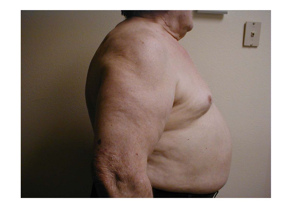
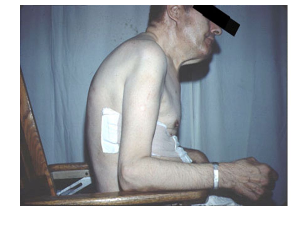
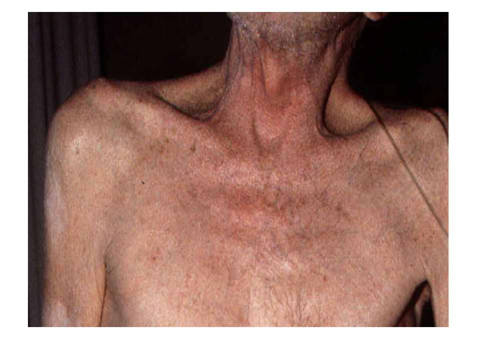
ΧΡΟΝΙΑ ΑΠΟΦΡΑΚΤΙΚΗ ΠΝΕΥΜΟΝΟΠΑΘΕΙΑ ( χρόνια βρογχίτιδα, πνευμονικό εμφύσημα)
Παρεμβολή αντιστάσεων στη ροή του αέρα, που δημιουργεί «απόφραξη» στην εκπνοή Μόνιμη αύξηση του μεγέθους των αεροφορών χώρων λόγω λέπτυσης και καταστροφής του τοιχώματός των κυψελίδων BLUE BLOATERS PINK PUFFERS

42
Βρογχικό άσθμα Είναι αποφρακτική πνευμονοπάθεια που χαρακτηρίζεται από εκτεταμένη και διάχυτη στένωση των αεραγωγών(βρογχιολιων) σα συνέπεια υπερβολικής απάντησης σε ερεθίσματα. 3% του πληθυσμού Παιδιά, αγόρια>κορίτσια Αλλεργία, λοίμωξη, υπερ δραστηριότητα παρασυμπαθητικού
 σα συνέπεια υπερβολικής απάντησης σε ερεθίσματα. 3% του πληθυσμού. Παιδιά, αγόρια>κορίτσια. Αλλεργία, λοίμωξη, υπερ δραστηριότητα παρασυμπαθητικού.")
43
Καρκίνος του πνεύμονα

44
Ατελεκτασία Είναι σύνδρομο που παρατηρείται σε πολλές παθολογικές καταστάσεις και σαν μετεγχειρητική επιπλοκή( ατελής- έκταση) Υποδηλώνει ατελή έκταση του πνεύμονα ή ενός τμήματός του και σύμπτωση των κυψελίδων

45
Πνευμονική εμβολή Είναι η απόφραξη του κλάδου της πνευμονικής αρτηρίας από κάποιο έμβολο, όπως θρόμβος αίματος που αποσπάται από φλέβες των κάτω άκρων και από τις δεξιές κοιλότητες της καρδιάς ( αέρας, λίπος, νεοπλασματικά κύτταρα, μυελός οστών κτλ) Σε μικρή εμβολή σύμπτωμα είναι η ταχυκαρδία Σε μαζική εμβολή->θάνατος
 Σε μικρή εμβολή σύμπτωμα είναι η ταχυκαρδία. Σε μαζική εμβολή->θάνατος.")
46
Πνευμονία Είναι οξεία λοίμωξη του πνευμονικού παρεγχύματος ( κυψελίδες), ανεξάρτητα από την έκταση που καταλαμβάνει και τον αιτιολογικό παράγοντα.
, ανεξάρτητα από την έκταση που καταλαμβάνει και τον αιτιολογικό παράγοντα.")
47
πνευμοθώρακας Συλλογή αέρα μέσα στην κοιλότητα του υπεζωκότα, με αποτέλεσμα τη σύμπτυξη του πνευμονικού παρεγχύματος και την εμπόδιση της αναπνευστικής λειτουργίας. Κλειστός ή ανοικτός ιατρογενής Τραυματικός, αυτόματος

48
Πλευρίτιδα Φλεγμονή στον υπεζωκότα

Παρόμοιες παρουσιάσεις


 Πασχάλης Στειρόπουλος.>")


