Κατέβασμα παρουσίασης
Η παρουσίαση φορτώνεται. Παρακαλείστε να περιμένετε

1
Ανατομία και Βιομηχανική
dsfsf Κρανίο- Σπονδυλική Στήλη- Αυχενική Μοίρα dsfsf dsfsf Ελευθερία Θωμαΐδου, Pt

2
Μαθησιακά Αποτελέσματα
Αναγνώριση των οστών του κρανίου και της σπονδυλικής στήλης και εντοπισμός τους στον ανθρώπινο σκελετό Περιγραφή της λειτουργίας της Σπονδυλικής Στήλης Περιγραφή της ανατομίας της Σ.Σ(Α.Μ.Σ.Σ) και των μεσοσπονδύλιων δίσκων Αναγνώριση των παρακείμενων αρθρώσεων Αναγνώριση και εντοπισμός μυών και συνδέσμων Α.Μ.ΣΣ
 και των μεσοσπονδύλιων δίσκων. Αναγνώριση των παρακείμενων αρθρώσεων. Αναγνώριση και εντοπισμός μυών και συνδέσμων Α.Μ.ΣΣ.")
3
Κρανίο Σύνθετη οστική κατασκευή
Ο σκελετός της κεφαλής υποδιαιρείται σε : Εγκεφαλικό κρανίο Σπλαχνικό κρανίο (σκελετός προσώπου) Αποτελείται από πλατιά οστά Τα οστά του κρανίου συνδέονται μεταξύ τους με ραφές
 Αποτελείται από πλατιά οστά. Τα οστά του κρανίου συνδέονται μεταξύ τους με ραφές.")
4
Κρανιακό θόλος-εγκεφαλικό κρανίο
Τα 8 οστά που σχηματίζουν τον εγκεφαλικό θόλο είναι: Το μετωπιαίο 2 βρεγματικά 2 κροταφικά Το σφηνοειδές Το ηθμοειδές Το ινιακό οστό

5
Μετωπιαίο οστό Πρόσθιο τοίχωμα του κρανίου

6
Βρεγματικά Οστά Πλάγια τοιχώματα του κρανίου

7
Κροταφικά οστά Κατώτερο τμήμα του πλάγιου τοιχώματος του κρανίου
Στυλοειδής απόφυση Μαστοειδής απόφυση

8
Σφηνοειδές οστό Σχήμα πεταλούδας
Το μεγαλύτερο μέρος του μεσαίου τμήματος της βάσης του κρανίου Αρθρώνεται με όλα τα κρανιακά οστά.

9
Ηθμοειδές οστό Το βαθύτερο κρανιακό οστό
Η περιοχή μεταξύ των οφθαλμικών κόγχων και ρινικής κοιλότητας

10
Ινιακό Οστό Οπίσθιο τοίχωμα του κρανίου
Περιλαμβάνει οστικά οδηγά σημεία: Το ινιακό όγκωμα Τις άνω και κάτω αυχενικές γραμμές

11
Cranial Bones Parietal Ethmoid Sphenoid Temporal Occipital Frontal

12
Οστά προσωπικού κρανίου
14 οστά 6 ζεύγη οστών 2 ανεξάρτητα οστά

13
Οστά προσωπικού κρανίου
2 ρινικά 2 υπερώια 2 δακρυϊκά 2 ζυγωματικά 2ρινικές κόγχες 2 οστά άνω γνάθου Ύνιδα Κάτω γνάθος

14
Κροταφογναθική διάρθρωση
Η άρθρωση μεταξύ κροταφικού οστού και κάτω γνάθου

15
Σπονδυλική Στήλη Οι 33 περίπου σπόνδυλοι διαχωρίζονται σε 5 ομάδες:
Αυχενική Μοίρα x7 Θωρακική Μοίρα x12 Οσφυϊκή Μοίρα x5 Ιερό Οστό x5 Κόκκυγας x3-5

16
Λειτουργία Σπονδυλικής Στήλης
Κρατάει όρθιο το σώμα Υποστηρίζει θώρακα, τον αυχένα και κρανίο Ορθοστατικός έλεγχος: κορμός & κρανίο Σημείο πρόσφυσης για τους μυς κίνηση Προστατεύει το νωτιαίο μυελό Απορρόφηση Δονήσεων: σπονδυλικά σώματα και δίσκοι

17
Σπονδυλική Στήλη Αυχενική : μεγάλο ROM άρα και μεγάλος κίνδυνος τραυματισμού Θωρακική :περιορισμένο ROM, μέγιστη προστασία Οσφυϊκά : ισορροπία μεταξύ ROM και κίνδυνου τραυματισμού Ιερό οστό και Κόκκυγας: συνοστεωμένα τμήματα ROM – range of movement

18
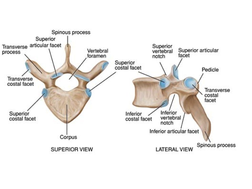
Τυπικός Σπόνδυλος Σώμα Αυχένας Σπονδυλικό τόξο πέταλο
Σπονδυλικός σωλήνας Εγκάρσια Απόφυση Άνω και κάτω αρθρική απόφυση Ακανθώδης απόφυση

20
Σπόνδυλοι

21
Όρια του Μεσοσπονδυλίου Τρήματος
IST/UH NMΣ2

22
Μεσοσπονδύλιος δίσκος
Από Α2-Ι1 Το μέγεθος είναι ανάλογο με το σπονδυλικό σώμα Συμβάλλει στην αύξηση του μήκους της ΣΣ κατά 20%

23
Μεσοσπονδύλιος Δίσκος
Ο μεσοσπονδύλιος δίσκος αποτελείται από ένα εξωτερικό ινώδη δακτύλιο που περιβάλλει ένα εσωτερικό πηκτοειδή πυρήνα Εκφυλιστικές αλλοιώσεις του ινώδους δακτυλίου είναι δυνατόν να οδηγήσουν σε δημιουργία πρόσπτωσης ( κήλης) του πηκτοειδούς πυρήνα
 του πηκτοειδούς πυρήνα.")
24
Αυχενικός και Οσφυϊκός Δίσκος
Distinctly different to the lumbar disc. Not surprising considering the form and function of the disc in the cervical spine. Axial rotation is a major function of the cervical spine whereas load bearing is more important in the lumbar. Nucleus pupolsis constitutes 25% of cervical spine disc (in lumbar its 50%0. Less volume in CSP. NP much more fibro cartilaginous than gelationous in CSP. From 30 years old the NP becomes dehydrated. Some studies suggest that by the age of 40 it is impossible to herniate the CSP disc. Higher levels of collagen to resist tensile forces. Clefts appear in the lateral aspect of the disc with age and maybe early signs of pathological degeneration or signs of the shearing which occurs at this level. The clefts appear from the ages of 9-14 and coincide with the uncinate processes reaching their maximum height. However the more deeply penetrating transverse fissures which transect the disc may be the result of degenerative changes. Annulus Fibrosis: Significantly different orientation of the lamellae than the lumbar disc. – alar fibres and vertically orientated fibres ΙST/UH NMΣ2

25
Αυχενικός και Οσφυϊκός Δίσκος
Οσφυϊκός δίσκος Κύρια λειτουργία η απορρόφηση των φορτίων Μεγαλύτερη ποσότητα ζελατινώδους ουσίας Εκφυλίζεται μετά τα 30 Μεγαλύτερος Μεγαλύτερη ποσότητα κολλαγόνου για τα φορτία Αυχενικός δίσκος Κύρια λειτουργία η στροφή Μεγαλύτερη ποσότητα ινοχόνδρινου ιστού Εκφυλίζεται μετά τα 40 Μικρότερος ΙST/UH NMΣ2

26
Όρια του Μεσοσπονδυλίου Τρήματος
Πρόσθια: σπονδυλικό σώμα Άνωθεν: πέταλο σπονδύλου : Facet IST/UH NMΣ2

27
Από το κέντρο στη περιφέρεια
IST/UH NMΣ2

28
Αυχενικοί Σπόνδυλοι

29
Οστικά Χαρακτηριστικά Αυχενικών Σπονδύλων
Σπονδυλικό Σώμα Μηνοειδής ακρολοφία Εγκάρσιο τρήμα Αρθρική επιφάνεια αυχενικών l facet Σπονδυλικό τόξο Δισχιδής ακανθώδης απόφυση Οι 7 αυχενικοί σπόνδυλοι χαρακτηρίζονται από το μικρό τους μέγεθος και τν ύπαρξη ενός τρήματος σε κάθε εγκάρσια απόφυση Το σπονδυλικό σώμα έχει μικρό ύψος και τετράγωνο σχήμα, όταν το βλέπουμε από πάνω και εμφανίζει μία άνω κοίλη επιφάνεια και μία κάτω κυρτή επιφάνεια Κάθε εγκάρσια απόφυση έχει αυλακοειδές σχήμα και διαπερνάται από ένα στρόγγυλο εγκάρσιο τρήμα Η ακανθώδης απόφυση έχει μικρό μήκος και διχάζεται σε δύο κορυφές Το σπονδυλικό τρήμα έχει τριγωνικό σχήμα ΙST/UH NMΣ2

30
ΙST/UH NMΣ2

31
Ανατομία ΑΜΣΣ Ανώτερη ΑΜΣΣ: ινιακό – Α2 Κατώτερη ΑΜΣΣ: Α3-Α7
ΙST/UH NMΣ2

32
Αυχενικοί σπόνδυλοι Μικρό σώμα σε μέγεθος 1-7

33
Α1 Άτλαντας Α1 Πρόσθιο φύμα πρόσθιο τόξο βοθρίο του οδόντα
άνω και κάτω αρθρικές επιφάνειες οπίσθιο τόξο οπίσθιο φύμα εγκάρσιες αποφύσεις εγκάρσιο τρήμα Basics: C1 Has an anterior tubercle, articular facet for the dens, lateral masses, inferior& superior articular facets, posterior arch, posterior tubercle & transverse process Slightly convex anteriorly, the short flattened anterior arch of C1 (atlas) has an anterior tubercle in the midline, giving attachment to the ALL, either side of which is the attachment of the superior oblique part of longus colli. Posteriorly, a concave circular facet articulates with the dens (odontoid process) of C2 (axis). The superior and inferior borders give attachment to the anterior atlanto-occipital membrane and anterior longitudinal ligament respectively. Atlas (First Cervical Vertebra): Anterior Arch Anatomy Text Slightly convex anteriorly, the short flattened anterior arch of C1 (atlas) has an anterior tubercle in the midline, giving attachment to the anterior longitudinal ligament (image) , either side of which is the attachment of the superior oblique part of longus colli. Posteriorly, a concave circular facet articulates with the dens (odontoid process) of C2 (axis). The superior and inferior borders give attachment to the anterior atlanto-occipital membrane and anterior longitudinal ligament respectively. Atlas (First Cervical Vertebra): Anterior Tubercle Anatomy Text The anterior tubercle is situated in the midline, on the anterior aspect of the anterior arch, where it gives attachment to the anterior longitudinal ligament (image) . Atlas (First Cervical Vertebra): Articular Facet for Dens Anatomy Text The articular facet for the dens is a concave circular facet located on the posterior aspect of the anterior arch of C1 (atlas), where it serves as an articular facet for the dens (odontoid process) (image) of C2 (axis). Atlas (First Cervical Vertebra): Inferior Articular Facets Anatomy Text The inferior articular processes of C1 (atlas) are flat or slightly concave circular facets located on the inferior surface of each lateral mass and are orientated more obliquely to the transverse plane than the superior articular facets and face postero-medially. The inferior articular facets articulate with the superior articular facets of C2 (axis) at the atlanto-axial joints (image) , thereby permitting rotation of the head and circumduction around the dens of the axis. Atlas (First Cervical Vertebra): Lateral Masses Anatomy Text The lateral masses are ovoid masses whose long axes converge anteriorly. Each has superior and inferior surfaces that bear superior and inferior articular facets, respectively. On the medial surface of each lateral mass is a roughened area with vascular foramina and a tubercle to which the transverse band of the cruciform ligament (image) (transverse ligament of atlas) attaches. Rectus capitis anterior attaches to the anterior surface of the lateral mass Atlas (First Cervical Vertebra): Posterior Arch Anatomy Text The posterior arch of C1 (atlas) forms most of a ring that represents the pedicles and laminae of a typical cervical vertebra. Immediately behind the lateral mass, the posterior arch is grooved on its superior surface by the vertebral artery (image) and vein and first cervical spinal nerve. This groove may be converted into a foramen (arcuate foramen) by cartilaginous or bony tissue (posterior ponticle) that bridges the posterior aspect of the lateral mass and the posterior arch. The posterior tubercle represents a rudimentary spinous process and gives attachment to the ligamentum nuchae. The rectus capitis posterior minor (animation) muscles attach lateral to the posterior tubercle. The superior and inferior borders give attachment to the posterior atlanto-occipital membrane and ligament flava, respectively. Atlas (First Cervical Vertebra): Posterior Tubercle Anatomy Text The posterior tubercle is situated in the midline, on the posterior aspect of the posterior arch. The tubercle is a rudimentary spinous process that projects posteriorly and gives attachment to the ligamentum nuchae. Atlas (First Cervical Vertebra): Superior Articular Facets on Lateral Masses Anatomy Text The superior articular facets are concave, reniform facets located on the superior surface of each lateral mass that face superiorly and medially. Each facet is narrowed in the middle (kidney-shaped) and occasionally completely divided into larger anterior and smaller posterior parts. They articulate with the occipital condyles at the atlanto-occipital joints (image) , thereby permitting flexion and extension of the head. As with the lateral masses themselves, the long axes of the facets converge anteriorly. Atlas (First Cervical Vertebra): Transverse Processes Anatomy Text The transverse processes of C1 (atlas) are longer than in other cervical vertebrae (except those of C7) and act as long levers. The broad flat apex represents the posterior tubercle of a typical cervical vertebra. Close to the lateral mass is the foramen transversarium that transmits the vertebral vessels (image) and associated sympathetic plexus. Several muscles attach to the transverse processes of C1. Obliquus capitis superior (animation) and rectus capitis lateralis attach superiorly to the transverse processes, with the latter displaying a more anterior site of attachment than the former. Laterally, obliquus capitis inferior (image) attaches to the apex, and scalenus medius, splenius cervicis and levator scapulae (antero-posteriorly) attach to the inferior surface ΙST/UH NMΣ2
 has an anterior tubercle in the midline, giving attachment to the ALL, either side of which is the attachment of the superior oblique part of longus colli. Posteriorly, a concave circular facet articulates with the dens (odontoid process) of C2 (axis). The superior and inferior borders give attachment to the anterior atlanto-occipital membrane and anterior longitudinal ligament respectively. Atlas (First Cervical Vertebra): Anterior Arch Anatomy Text Slightly convex anteriorly, the short flattened anterior arch of C1 (atlas) has an anterior tubercle in the midline, giving attachment to the anterior longitudinal ligament (image) , either side of which is the attachment of the superior oblique part of longus colli. Posteriorly, a concave circular facet articulates with the dens (odontoid process) of C2 (axis). The superior and inferior borders give attachment to the anterior atlanto-occipital membrane and anterior longitudinal ligament respectively. Atlas (First Cervical Vertebra): Anterior Tubercle Anatomy Text The anterior tubercle is situated in the midline, on the anterior aspect of the anterior arch, where it gives attachment to the anterior longitudinal ligament (image) . Atlas (First Cervical Vertebra): Articular Facet for Dens Anatomy Text The articular facet for the dens is a concave circular facet located on the posterior aspect of the anterior arch of C1 (atlas), where it serves as an articular facet for the dens (odontoid process) (image) of C2 (axis). Atlas (First Cervical Vertebra): Inferior Articular Facets Anatomy Text The inferior articular processes of C1 (atlas) are flat or slightly concave circular facets located on the inferior surface of each lateral mass and are orientated more obliquely to the transverse plane than the superior articular facets and face postero-medially. The inferior articular facets articulate with the superior articular facets of C2 (axis) at the atlanto-axial joints (image) , thereby permitting rotation of the head and circumduction around the dens of the axis. Atlas (First Cervical Vertebra): Lateral Masses Anatomy Text The lateral masses are ovoid masses whose long axes converge anteriorly. Each has superior and inferior surfaces that bear superior and inferior articular facets, respectively. On the medial surface of each lateral mass is a roughened area with vascular foramina and a tubercle to which the transverse band of the cruciform ligament (image) (transverse ligament of atlas) attaches. Rectus capitis anterior attaches to the anterior surface of the lateral mass. Atlas (First Cervical Vertebra): Posterior Arch Anatomy Text The posterior arch of C1 (atlas) forms most of a ring that represents the pedicles and laminae of a typical cervical vertebra. Immediately behind the lateral mass, the posterior arch is grooved on its superior surface by the vertebral artery (image) and vein and first cervical spinal nerve. This groove may be converted into a foramen (arcuate foramen) by cartilaginous or bony tissue (posterior ponticle) that bridges the posterior aspect of the lateral mass and the posterior arch. The posterior tubercle represents a rudimentary spinous process and gives attachment to the ligamentum nuchae. The rectus capitis posterior minor (animation) muscles attach lateral to the posterior tubercle. The superior and inferior borders give attachment to the posterior atlanto-occipital membrane and ligament flava, respectively. Atlas (First Cervical Vertebra): Posterior Tubercle Anatomy Text The posterior tubercle is situated in the midline, on the posterior aspect of the posterior arch. The tubercle is a rudimentary spinous process that projects posteriorly and gives attachment to the ligamentum nuchae. Atlas (First Cervical Vertebra): Superior Articular Facets on Lateral Masses Anatomy Text The superior articular facets are concave, reniform facets located on the superior surface of each lateral mass that face superiorly and medially. Each facet is narrowed in the middle (kidney-shaped) and occasionally completely divided into larger anterior and smaller posterior parts. They articulate with the occipital condyles at the atlanto-occipital joints (image) , thereby permitting flexion and extension of the head. As with the lateral masses themselves, the long axes of the facets converge anteriorly. Atlas (First Cervical Vertebra): Transverse Processes Anatomy Text The transverse processes of C1 (atlas) are longer than in other cervical vertebrae (except those of C7) and act as long levers. The broad flat apex represents the posterior tubercle of a typical cervical vertebra. Close to the lateral mass is the foramen transversarium that transmits the vertebral vessels (image) and associated sympathetic plexus. Several muscles attach to the transverse processes of C1. Obliquus capitis superior (animation) and rectus capitis lateralis attach superiorly to the transverse processes, with the latter displaying a more anterior site of attachment than the former. Laterally, obliquus capitis inferior (image) attaches to the apex, and scalenus medius, splenius cervicis and levator scapulae (antero-posteriorly) attach to the inferior surface. ΙST/UH NMΣ2.")
34
Άτλας – Α1 Υποστηρίζει το κεφάλι
Δεν έχει σώμα, ούτε ακανθώδη απόφυση( a ring) Αρθρώνεται με το ινιακό οστό και σχηματίζει την ατλαντοινιακή άρθρωση.
 Αρθρώνεται με το ινιακό οστό και σχηματίζει την ατλαντοινιακή άρθρωση.")
35
Α2 - Άξονας Α2: Οδόντας μακριά ακανθώδη απόφυση εγκάρσιο τρήμα
Basics: The dens (odontoid process) is a conical projection that arises from the superior surface of the vertebral body of C2 The spinous process of C2 (axis) is a large, posteriorly orientated process associated with a broad inferiorly concave base and bifid apex The inferior articular facets of the axis are situated on its inferior articular processes. These facets are oval, slightly concave in shape and face antero-inferiorly for articulation with the superior articular facets of C3 at the facet joint. The superior articular facets of C2 (axis) are slightly convex in shape and project supero-laterally from the superior surface of the pedicles and the anterior part of the transverse processes. The superior articular facets are covered with hyaline cartilage and articulate with the inferior articular facets of C1 (atlas) at the lateral atlanto-axial joints, (image) which permit forward and backward gliding of the C1 lateral masses during rotation on C2. Laterally, the laminae of C2 (axis) are thick, strong and concave. They run postero-medially, thereby forming a connection between the pedicles and the spinous process. Axis (Second Cervical Vertebra): Articular Facet for Anterior Arch of Atlas Anatomy Text On the anterior aspect of the dens (odontoid process) lies an ovoid, vertically orientated facet. It is covered with hyaline cartilage and articulates with the facet on the posterior aspect of the anterior arch of C1 (atlas) at the median atlanto-axial joint. Axis (Second Cervical Vertebra): Bifid Spinous Process Anatomy Text The spinous process of C2 (axis) is a large, posteriorly orientated process associated with a broad inferiorly concave base and bifid apex. Obliquus capitis inferior attaches to the lateral surfaces anteriorly and rectus capitis posterior major (image) posteriorly. Multifidus (animation) , semispinalis cervicis and spinalis cervicis attach to the inferior concavity antero-posteriorly and interspinales attaches near the apex. The ligamentum nuchae attaches to the apical notch of the spinous process and the interspinous ligament attaches to the inferior margin, in the midline. Axis (Second Cervical Vertebra): Body Anatomy Text The body of C2 (axis) consists of partly fused centra of C1 (atlas) and C2 and a rudimentary intervertebral disc between. A deep depression on each side of the anterior surface gives attachment to longus colli. The inferiorly projecting anterior border gives attachment to the anterior longitudinal ligament (image) , whereas the flat or slightly concave posterior surface gives attachment to the posterior longitudinal ligament, the deep and superficial laminae of the tectorial membrane and the inferior band of the cruciform ligament (image) (from the lower border of the transverse band of the cruciform ligament - transverse ligament of atlas). The inferior surface has a thin covering of hyaline cartilage, the ‘end-plate’, to which the adjacent intervertebral disc is attached Axis (Second Cervical Vertebra): Dens (Syn. Odontoid Process) Anatomy Text Jump to Clinical Pathology Text The dens (odontoid process) is a conical projection that arises from the superior surface of the vertebral body of C2 (axis) (image) . It may be tilted posteriorly, anteriorly or laterally with respect to the vertebral body. The posterior surface is grooved for the transverse band of the cruciform ligament (transverse ligament of atlas), above which is the head. The anterior surface has an ovoid facet for articulation with the facet on the posterior aspect of the anterior arch of C1 (atlas). The apical ligament attaches to the pointed apex whereas the alar ligaments attach to ovoid flattenings on the postero-lateral aspect of the apex of the dens. At the junction of the body and vertebral arch are large ovoid flat or slightly convex superior articular facets for articulation with the inferior articular facets of C1 (atlas) at the atlanto-axial joint (image) . Axis (Second Cervical Vertebra): Inferior Articular Facets Anatomy Text The inferior articular facets of the axis are situated on its inferior articular processes. These facets are oval, slightly concave in shape and face antero-inferiorly for articulation with the superior articular facets of C3 at the zygapophyseal joint (image) . Axis (Second Cervical Vertebra): Laminae Anatomy Text Laterally, the laminae of C2 (axis) are thick, strong and concave. They run postero-medially, thereby forming a connection between the pedicles and the spinous process. Ligamentum flava pass from the superior and inferior borders to adjacent borders of C1 and C3, respectively. Axis (Second Cervical Vertebra): Superior Articular Facets for Inferior Facets of Atlas Anatomy Text Axis (Second Cervical Vertebra): Transverse Processes Anatomy Text The transverse processes of C2 (axis) (image) , which contain the foramen transversarium, are infero-lateral projections arising from the pedicle-laminar junction and lateral aspect of the interarticular area of the pedicle. The rounded apex is equivalent to the posterior tubercle of a typical cervical vertebrae. The muscles attaching to the transverse processes include scalenus medius, levator scapulae (animation) and splenius cervicis, which attach to the tip antero-posteriorly and the intertransversarii muscles, which attach to the superior and inferior surfaces. Α2: Οδόντας μακριά ακανθώδη απόφυση εγκάρσιο τρήμα άνω/κάτω αρθρικές αποφύσεις τόξο. ΙST/UH NMΣ2
 is a conical projection that arises from the superior surface of the vertebral body of C2. The spinous process of C2 (axis) is a large, posteriorly orientated process associated with a broad inferiorly concave base and bifid apex. The inferior articular facets of the axis are situated on its inferior articular processes. These facets are oval, slightly concave in shape and face antero-inferiorly for articulation with the superior articular facets of C3 at the facet joint. The superior articular facets of C2 (axis) are slightly convex in shape and project supero-laterally from the superior surface of the pedicles and the anterior part of the transverse processes. The superior articular facets are covered with hyaline cartilage and articulate with the inferior articular facets of C1 (atlas) at the lateral atlanto-axial joints, (image) which permit forward and backward gliding of the C1 lateral masses during rotation on C2. Laterally, the laminae of C2 (axis) are thick, strong and concave. They run postero-medially, thereby forming a connection between the pedicles and the spinous process. Axis (Second Cervical Vertebra): Articular Facet for Anterior Arch of Atlas Anatomy Text. On the anterior aspect of the dens (odontoid process) lies an ovoid, vertically orientated facet. It is covered with hyaline cartilage and articulates with the facet on the posterior aspect of the anterior arch of C1 (atlas) at the median atlanto-axial joint. Axis (Second Cervical Vertebra): Bifid Spinous Process Anatomy Text. The spinous process of C2 (axis) is a large, posteriorly orientated process associated with a broad inferiorly concave base and bifid apex. Obliquus capitis inferior attaches to the lateral surfaces anteriorly and rectus capitis posterior major (image) posteriorly. Multifidus (animation) , semispinalis cervicis and spinalis cervicis attach to the inferior concavity antero-posteriorly and interspinales attaches near the apex. The ligamentum nuchae attaches to the apical notch of the spinous process and the interspinous ligament attaches to the inferior margin, in the midline. Axis (Second Cervical Vertebra): Body Anatomy Text. The body of C2 (axis) consists of partly fused centra of C1 (atlas) and C2 and a rudimentary intervertebral disc between. A deep depression on each side of the anterior surface gives attachment to longus colli. The inferiorly projecting anterior border gives attachment to the anterior longitudinal ligament (image) , whereas the flat or slightly concave posterior surface gives attachment to the posterior longitudinal ligament, the deep and superficial laminae of the tectorial membrane and the inferior band of the cruciform ligament (image) (from the lower border of the transverse band of the cruciform ligament - transverse ligament of atlas). The inferior surface has a thin covering of hyaline cartilage, the ‘end-plate’, to which the adjacent intervertebral disc is attached. Axis (Second Cervical Vertebra): Dens (Syn. Odontoid Process) Anatomy Text Jump to Clinical Pathology Text. The dens (odontoid process) is a conical projection that arises from the superior surface of the vertebral body of C2 (axis) (image) . It may be tilted posteriorly, anteriorly or laterally with respect to the vertebral body. The posterior surface is grooved for the transverse band of the cruciform ligament (transverse ligament of atlas), above which is the head. The anterior surface has an ovoid facet for articulation with the facet on the posterior aspect of the anterior arch of C1 (atlas). The apical ligament attaches to the pointed apex whereas the alar ligaments attach to ovoid flattenings on the postero-lateral aspect of the apex of the dens. At the junction of the body and vertebral arch are large ovoid flat or slightly convex superior articular facets for articulation with the inferior articular facets of C1 (atlas) at the atlanto-axial joint (image) . Axis (Second Cervical Vertebra): Inferior Articular Facets Anatomy Text. The inferior articular facets of the axis are situated on its inferior articular processes. These facets are oval, slightly concave in shape and face antero-inferiorly for articulation with the superior articular facets of C3 at the zygapophyseal joint (image) . Axis (Second Cervical Vertebra): Laminae Anatomy Text. Laterally, the laminae of C2 (axis) are thick, strong and concave. They run postero-medially, thereby forming a connection between the pedicles and the spinous process. Ligamentum flava pass from the superior and inferior borders to adjacent borders of C1 and C3, respectively. Axis (Second Cervical Vertebra): Superior Articular Facets for Inferior Facets of Atlas Anatomy Text. Axis (Second Cervical Vertebra): Transverse Processes Anatomy Text. The transverse processes of C2 (axis) (image) , which contain the foramen transversarium, are infero-lateral projections arising from the pedicle-laminar junction and lateral aspect of the interarticular area of the pedicle. The rounded apex is equivalent to the posterior tubercle of a typical cervical vertebrae. The muscles attaching to the transverse processes include scalenus medius, levator scapulae (animation) and splenius cervicis, which attach to the tip antero-posteriorly and the intertransversarii muscles, which attach to the superior and inferior surfaces. Α2: Οδόντας. μακριά ακανθώδη απόφυση. εγκάρσιο τρήμα. άνω/κάτω αρθρικές αποφύσεις. τόξο. ΙST/UH NMΣ2.")
36
Άξονας – Α2 Πολύ μικρό σώμα
Ο Οδόντας λειτουργεί σαν ένας άξονας που επιτρέπει στον Α1 σπόνδυλο και την κεφαλή να στρέφονται δεξιά και αριστερά γύρω από αυτόν Ο Α1 και Α2 σχηματίζουν την Ατλαντοαξονική άρθρωση

37
Αυχενικός Σπόνδυλος

38
Α7 Α7: Ο Α7 είναι μεγαλύτερος από τους προηγούμενους σπονδύλους και εμφανίζει χαρακτηριστικά παρόμοια με αυτά των θωρακικών σπονδύλων: Μεγαλύτερο σπονδυλικό σώμα Μικρότερο σπονδυλικό τρήμα Οι κάτω αρθρικές αποφύσεις έχουν μεγαλύτερή πρόσθια γωνίωση Οι άνω αρθρικές αποφύσεις έχουν μεγαλύτερη οπίσθια γωνίωση The seventh cervical vertebra (animation) (C7) is larger than the preceding cervical vertebrae and displays characteristics similar to those of the thoracic vertebrae; -Larger vertebral body -Smaller vertebral foramen -Pedicles directed more posteriorly rather than laterally -Inferior articular facets directed more anteriorly than inferiorly -Superior articular facets more posteriorly than superiorly C7 has a broad vertebral body and triangular vertebral foramen (image) . The vertebral foramen is bound by the vertebral body anteriorly, pedicles laterally and laminae postero-laterally. It contains the spinal cord (animation) surrounded by the meninges and associated vessels. The anterior surface of the vertebral body is convex and has a downward projecting lip to which the anterior longitudinal ligament (image) attaches. The posterior surface is concave and the superior surface is saddle-shaped with lateral elevations known as the uncinate processes which articulate with reciprocal articular facets located on the inferior (concave) surface of the vertebra above. These articulations are known as the ‘uncovertebral joints’ or ‘joints of Luschka’ The pedicles project postero-laterally and are notched on their superior and inferior surfaces by the superior and inferior vertebral notches, respectively; they contribute to the formation of the boundaries of the intervertebral foramina. The thin, curved laminae project postero-medially and have thinner superior and thicker inferior borders. The spinous process of C7 projects from the junction of the laminae. It is atypical in that it resembles a thoracic spinous process; it is long, non-bifid, and ends in a prominent tubercle, which is easily palpable (image) . The thick transverse processes end laterally in anterior and prominent posterior tubercles joined by a thin costotransverse bar. Within each transverse process is a small (and sometimes absent) foramen transversarium. Unlike the other cervical vertebrae the foramina transversaria of C7 do not transmit the vertebral artery, although they frequently transmit vertebral veins. At the junction of the pedicles and laminae project large superior and inferior articular processes, on which are the slightly convex superior and slightly concave inferior articular facets. The superior articular facets face postero-superiorly and the inferior articular facets face antero-inferiorly. The superior and inferior articular processes from C2 (inferior articular process) to C7 form the articular pillar of the cervical spine. It is palpable 2-3cm from the cervical spinous processes ΙST/UH NMΣ2
 (C7) is larger than the preceding cervical vertebrae and displays characteristics similar to those of the thoracic vertebrae; -Larger vertebral body. -Smaller vertebral foramen. -Pedicles directed more posteriorly rather than laterally. -Inferior articular facets directed more anteriorly than inferiorly. -Superior articular facets more posteriorly than superiorly. C7 has a broad vertebral body and triangular vertebral foramen (image) . The vertebral foramen is bound by the vertebral body anteriorly, pedicles laterally and laminae postero-laterally. It contains the spinal cord (animation) surrounded by the meninges and associated vessels. The anterior surface of the vertebral body is convex and has a downward projecting lip to which the anterior longitudinal ligament (image) attaches. The posterior surface is concave and the superior surface is saddle-shaped with lateral elevations known as the uncinate processes which articulate with reciprocal articular facets located on the inferior (concave) surface of the vertebra above. These articulations are known as the ‘uncovertebral joints’ or ‘joints of Luschka’ The pedicles project postero-laterally and are notched on their superior and inferior surfaces by the superior and inferior vertebral notches, respectively; they contribute to the formation of the boundaries of the intervertebral foramina. The thin, curved laminae project postero-medially and have thinner superior and thicker inferior borders. The spinous process of C7 projects from the junction of the laminae. It is atypical in that it resembles a thoracic spinous process; it is long, non-bifid, and ends in a prominent tubercle, which is easily palpable (image) . The thick transverse processes end laterally in anterior and prominent posterior tubercles joined by a thin costotransverse bar. Within each transverse process is a small (and sometimes absent) foramen transversarium. Unlike the other cervical vertebrae the foramina transversaria of C7 do not transmit the vertebral artery, although they frequently transmit vertebral veins. At the junction of the pedicles and laminae project large superior and inferior articular processes, on which are the slightly convex superior and slightly concave inferior articular facets. The superior articular facets face postero-superiorly and the inferior articular facets face antero-inferiorly. The superior and inferior articular processes from C2 (inferior articular process) to C7 form the articular pillar of the cervical spine. It is palpable 2-3cm from the cervical spinous processes. ΙST/UH NMΣ2.")
39
Θωρακικός Σπόνδυλος 12 θωρακικοί σπόνδυλοι αρθρώνονται με τις πλευρές
Κάθε τυπικός θωρακικός σπόνδυλος έχει σε κάθε πλευρά του σώματος δύο ημιγλήνια για τν άρθρωση με την πλευρά. Οι κατώτεροι θωρακικοί σπόνδυλοι μοιάζουν με τους οσφυικούς TP – transverse process SP – spinous process

40
Thoracic Vertebrae

41
Facet Οι διαρθρώσεις μεταξύ άνω και κάτω αρθρικών αποφύσεων ονομάζονται ζυγοαποφυσιακές αρθρώσεις ΙST/UH NMΣ2

42
Αυχενικές Ζυγαποφυσιακές Αρθρώσεις-Facet
Χαλαρός αρθρικός θύλακας Nεύρωση: Μέσος κλάδος των ραχιαίων κλάδων των νωτιαίων νεύρων Λειτουργία: Ο προσανατολισμός των Facet διευκολύνει τις κινήσεις στο οβελιαίο επίπεδο Facet orientation facilitates sagittal plane movements, meaning that rotation and lateral flexion are always a coupled movement. Joint capsule is lax, facilitating great amount of movements –Capsule partly formed by ligamentum flavum anteriorly and posteriorly capsule is thin. There is often a large triangular fat pad as the lower posterior portion of the joint which is enclosed by the insertions of deep multifidus. ΙST/UH NMΣ2

43
Μηνοειδής Ακρολοφία – Αρθρώσεις του Luschka
Άρθρωση: Η μηνοειδής ακρολοφία του κάτω σπονδύλου αρθρώνεται με τον ανώτερο σπόνδυλο. Οι αρθρώσεις αυτές λέγονται αρθρώσεις του Luschka’ λειτουργία: Ελαττώνει το μεσάρθριο διάστημα, αυξάνει την πλάγια σταθερότητα The anterior surface of the vertebral body is convex and has a downward projecting lip to which the ALL attaches. The posterior surface is concave and the superior surface is saddle-shaped with lateral elevations known as the uncinate processes which articulate with reciprocal articular facets located on the inferior (concave) surface of the vertebra above. These articulations are known as the ‘uncovertebral joints’ or ‘joints of Luschka’. Some debate as to whether these are true joints or fissures which develop in the lateral aspect of the disc. ΙST/UH NMΣ2
 surface of the vertebra above. These articulations are known as the ‘uncovertebral joints’ or ‘joints of Luschka’. Some debate as to whether these are true joints or fissures which develop in the lateral aspect of the disc. ΙST/UH NMΣ2.")
44
ΙST/UH NMΣ2

45
Αυχενικός σύνδεσμος ΙST/UH NMΣ2

46
Αυχενικοί Σύνδεσμοι Αυχενικός Σύνδεσμος: Επακάνθιος Σύνδεσμος
Πυκνή τραπεζοειδής, τριγωνική μεμβράνη δύο πτυχών Ελαστική & ινώδης Εκφύεται από το έξω ινιακό όγκωμα και καταφύεται στην ακαθώδη απόφυση του Α7 Επίσης προσφύεται στο έσω χείλος των δισχιδών ακανθωδών αποφύσεων όλων των αυχενικών σπονδύλων Προσφέρει πρόσφυση για αυχενικούς μυς Επακάνθιος Σύνδεσμος The ligamentum nuchae is a dense, bilaminar, triangular midline fibroelastic intermuscular septum. It extends from the external occipital protuberance to the spine of C7 and attaches to the median part of the external occipital crest, the posterior tubercle of C1 and the medial aspect of the cervical bifid spines. It provides attachment for cervical muscles and is distinct from the supraspinous and interspinous ligaments, although appears to largely replace this in cervical spine. ΙST/UH NMΣ2

47
Αυχενικοί Σύνδεσμοι Πρόσθιος επιμήκης: Οπίσθιος επιμήκης
Α1 - ιερό Οπίσθιος επιμήκης Α2 - Ι1 Ωχρός Σύνδεσμος Α1/2 έως Ο4/5 από τόξο σε τόξο Μεσεγκάρσιος Μεσακάνθιος Επακάνθιος Anterior Longitudinal Ligament Anatomy Text The anterior longitudinal ligament is a strong band supporting the anterior aspect of the vertebral column. It is narrower superiorly (image) (4-5mm) and broader inferiorly (image) (20-25mm). It extends from the anterior tubercle of C1 to the pelvic surface of the upper sacrum. It is between 1-2mm thick and consists of three dense layers of collagen fibers. The superficial layer extends over three or four vertebrae; the intermediate layer over two or three and the deep layer consists of laterally short fibers connecting adjacent vertebrae. The fibers are strongly attached to the intervertebral margins, but loosely to the bodies. Superiorly, the ligament is continuous with the anterior atlanto-occipital membrane. Posterior Longitudinal Ligament Anatomy Text The posterior longitudinal ligament is a strong band supporting the posterior aspect of the vertebral column. It is wider superiorly (10-15mm) and narrower and denticulate inferiorly (6-8mm over the bodies; 10-15mm over the discs). It extends from the posterior surface of the body of C2 to the posterior surface of S1. It is 1-1.4mm thick and consists of two dense layers of collagen fibers. The superficial fibers extend over three or four vertebrae and the deeper ones pass between adjacent vertebrae. The fibers attach to the intervertebral discs and the adjacent margins of the vertebral bodies. Between attachments they are separated from the bodies by basivertebral veins and their communications with the anterior internal vertebral venous plexus. Superiorly, the ligament is continuous with the tectorial membrane. Ligamentum Flava Anatomy Text The ligamentum flava connects the laminae of adjacent vertebrae. The first arises at C1/C2 and the last at the level of L4/L5 (L5/S1 in a lumbarized sacrum). They pass postero-inferiorly from the anterior aspect of the inferior border of the laminae above to the posterior aspect of the superior border of the laminae below. Laterally, the ligaments extend as far as the zygapophyseal joint capsules. Medially, their borders meet at the root of the spinous process and may be partially fused leaving gaps for the passage of veins connecting the internal and posterior external vertebral venous plexus. The predominant tissue is yellow elastic tissue to permit and control spinal flexion and prevent abrupt limitation, they also aid the return to the erect posture and prevent redundant folds projecting into the vertebral foramen. The ligaments are broad, thin and long in the cervical region, becoming thicker as they descend the vertebral column. Intertransverse ligt are, generally, insignificant bands connecting adjacent transverse processes. In the cervical region, they consist of irregular fibers largely replaced by intertransversarii muscles. In the thoracic region, they intimately blend with adjacent muscles and in the lumbar region they are thin and membranous. ΙST/UH NMΣ2
 (4-5mm) and broader inferiorly (image) (20-25mm). It extends from the anterior tubercle of C1 to the pelvic surface of the upper sacrum. It is between 1-2mm thick and consists of three dense layers of collagen fibers. The superficial layer extends over three or four vertebrae; the intermediate layer over two or three and the deep layer consists of laterally short fibers connecting adjacent vertebrae. The fibers are strongly attached to the intervertebral margins, but loosely to the bodies. Superiorly, the ligament is continuous with the anterior atlanto-occipital membrane. Posterior Longitudinal Ligament Anatomy Text. The posterior longitudinal ligament is a strong band supporting the posterior aspect of the vertebral column. It is wider superiorly (10-15mm) and narrower and denticulate inferiorly (6-8mm over the bodies; 10-15mm over the discs). It extends from the posterior surface of the body of C2 to the posterior surface of S1. It is 1-1.4mm thick and consists of two dense layers of collagen fibers. The superficial fibers extend over three or four vertebrae and the deeper ones pass between adjacent vertebrae. The fibers attach to the intervertebral discs and the adjacent margins of the vertebral bodies. Between attachments they are separated from the bodies by basivertebral veins and their communications with the anterior internal vertebral venous plexus. Superiorly, the ligament is continuous with the tectorial membrane. Ligamentum Flava Anatomy Text. The ligamentum flava connects the laminae of adjacent vertebrae. The first arises at C1/C2 and the last at the level of L4/L5 (L5/S1 in a lumbarized sacrum). They pass postero-inferiorly from the anterior aspect of the inferior border of the laminae above to the posterior aspect of the superior border of the laminae below. Laterally, the ligaments extend as far as the zygapophyseal joint capsules. Medially, their borders meet at the root of the spinous process and may be partially fused leaving gaps for the passage of veins connecting the internal and posterior external vertebral venous plexus. The predominant tissue is yellow elastic tissue to permit and control spinal flexion and prevent abrupt limitation, they also aid the return to the erect posture and prevent redundant folds projecting into the vertebral foramen. The ligaments are broad, thin and long in the cervical region, becoming thicker as they descend the vertebral column. Intertransverse ligt are, generally, insignificant bands connecting adjacent transverse processes. In the cervical region, they consist of irregular fibers largely replaced by intertransversarii muscles. In the thoracic region, they intimately blend with adjacent muscles and in the lumbar region they are thin and membranous. ΙST/UH NMΣ2.")
48
Σύνδεσμοι Ανώτερης Αυχενικής Μοίρας
Πτερυγοειδείς Σύνδεσμοι Είναι παχιές ταινίες παράλληλα προσανατολισμένων κολλαγόνων ινών που εκτείνονται από το οδόντα του άξονα στους ινιακούς κονδύλους. Περιορίζει την αντίθετη πλάγια κάμψη Alar Ligaments Anatomy Text The alar ligaments are thick cords of parallel collagen fibers extending horizontally and laterally from ovoid regions on the postero-lateral aspect of the apex of the dens to roughened areas on the medial sides of the occipital condyles. Most individuals also have an antero-inferior band, which attaches to the lateral mass of C1 anterior to the transverse atlantal ligament, with occasionally a few fibers passing from the dens to the anterior arch of C1. The ligaments limit atlanto-axial rotation, however slight upward movement of the axis during rotation helps permit a wider range of movement by reducing tension in the ligaments. ΙST/UH NMΣ2

49
Σύνδεσμοι Ανώτερης Αυχενικής Μοίρας
Κορυφαίος Σύνδεσμος Είναι μια λεπτή ταινία που εκτείνεται από την κορυφή του οδόντα στο πρόσθιο χείλος του ινιακού τρήματος ανάμεσα από του πτερυγοειδείς συνδέσμους. Εμποδίζει την απομάκρυνση Α0-Α1 Apical Ligament of the Dens Anatomy Text The apical ligament of the dens is a slender band, fanning out from the apex of the dens to the anterior margin of the foramen magnum between the alar ligaments. It is positioned with the anterior atlanto-occipital membrane anteriorly and the superior band of the cruciform ligament posteriorly, and is separated from both by pads of fatty tissue, but blends with their attachments at the foramen magnum. It is said to represent the cranial continuation of the notochord and its sheath. ΙST/UH NMΣ2

50
Εγκάρσιος σύνδεσμος Ισχυρή δεσμίδα
Προσφύεται μεταξύ των 2 πλάγιων ογκωμάτων του άτλαντα Συγκρατεί τον οδόντα στο οστικό δαχτυλίδι του άτλαντα Η ακεραιότητα του είναι ζωτικής σημασίας Περιορίζει τη κάμψη και τη πρόσθια μετατόπιση του άτλαντα ΙST/UH NMΣ2

51
Εγκάρσιος σύνδεσμος ΙST/UH NMΣ2

52
Ανώτερη Μοίρα Ποιοι σύνδεσμοι βοηθούν τον έλεγχο κινήσεων στην άνω αυχενική; ΙST/UH NMΣ2

53
Σπονδυλικές Αρτηρίες Branches from the subclavian artery at the level of T1 Ascends between Longus Colli & scalenus anterior Passes through the foramen of C6 with a branch from the inferior sympathetic ganglion and vertebral venous plexus Ascends vertically through foramina transversaria of C5 to C2 From the foramen transversarium of C2 (axis) the VA passes laterally to enter the foramen transversarium of C1 (atlas). Each vertebral artery then lies in the groove on the superior surface of the posterior arch of C1 and enters the vertebral canal below the inferior border of the posterior atlanto-occipital membrane. The vertebral artery subsequently pierces the dura and arachnoid mater, ascends into the skull through the foramen magnum. ΙST/UH NMΣ2
 the VA passes laterally to enter the foramen transversarium of C1 (atlas). Each vertebral artery then lies in the groove on the superior surface of the posterior arch of C1 and enters the vertebral canal below the inferior border of the posterior atlanto-occipital membrane. The vertebral artery subsequently pierces the dura and arachnoid mater, ascends into the skull through the foramen magnum. ΙST/UH NMΣ2.")
54
Quiz Πως αλλιώς λέγεται ο Α1; Πως αλλιώς λέγεται ο Α2; ΙST/UH NMΣ2

55
Quiz Πως λέγονται οι αρθρώσεις που σχηματίζονται από την μηνοειδή ακρολοφία και το σώμα του ανώτερου σπονδύλου; ΙST/UH NMΣ2

56
Quiz Ποιός είναι αυτός ο σύνδεσμoς; ΙST/UH NMΣ2

Παρόμοιες παρουσιάσεις