Κατέβασμα παρουσίασης
Η παρουσίαση φορτώνεται. Παρακαλείστε να περιμένετε

1
Μυκωτικά Ανευρύσματα – Μόλυνση Ενδοπρόθεσης Κωνσταντίνος Γ. Μουλακάκης
Κωνσταντίνος Γ. Μουλακάκης Επιμελητής A Αγγειοχειρουργός, Αγγειοχειρουργική Κλινική, “ Aττικόν” Νοσοκομείο 2015

2
"infected aneurysm" has gradually replaced the original designation "mycotic aneurysm“

3
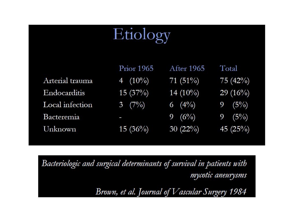
Μυκωτικά Ανευρύσματα – Αιτιολογία και Παράγοντες Κινδύνου
Μυκωτικά Ανευρύσματα – Αιτιολογία και Παράγοντες Κινδύνου Βακτηριακή Ενδοκαρδίτιδα (σηπτικά έμβολα) Αρτηριακό Τραύμα, ή τραύμα του ενδοθηλίου (Τοξικομανείς, Στεφανιογραφίες, ΧΝΑ) Φλεγμονή, Μικροβιαιμία (Ουρολοίμωξη, Πνευμονία, Εκκολπωματίτιδα) Διασπορά από Φλεγμονή περιξ της αορτής (σπονδυλίτιδα, γαστρεντεριτιδα με προσβολή παραορτικών λεμφαδένων) Διαταραχές ανοσοποιητικού συστήματος (70%) Αθηροσκλήρωση (αποικισμός πλακών από μικρόβια)
 Αρτηριακό Τραύμα, ή τραύμα του ενδοθηλίου (Τοξικομανείς, Στεφανιογραφίες, ΧΝΑ) Φλεγμονή, Μικροβιαιμία (Ουρολοίμωξη, Πνευμονία, Εκκολπωματίτιδα) Διασπορά από Φλεγμονή περιξ της αορτής (σπονδυλίτιδα, γαστρεντεριτιδα με προσβολή παραορτικών λεμφαδένων) Διαταραχές ανοσοποιητικού συστήματος (70%) Αθηροσκλήρωση (αποικισμός πλακών από μικρόβια)")
5
Ο ρόλος της ενδαγγειακής αποκατάστασης στη θεραπεία των Μυκωτικών ανευρυσμάτων

6
Εμπλεκόμενοι Μικροοργανισμοί
Θετικές Καλλιέργειες ≈ 62 % Staphylococcus sp. 20 % Salmonella % Streptococcus sp % Other % (Pseudomonas aeruginosa, E coli, Enterobacterium faecium, Prot. mirabilis, Serratia sonticola, Bacteroides fr., Bacillus cereus, Tuberculous species, Listeria monocytogenes, Coxiella bruneti, Candida albicans) ↑f Consistently the most common identified microorganisms In the Asian population, 3 characteristics are apparent: (1) most patients are immunocompromised; (2) patients present late in the course of disease; and (3) Salmonella is usually responsible. Today, the majority of reported organisms found in aortic infections are gram-positive cocci, accounting for approximately 60% of reported cases of aortitis.[2,3,5] Staphylococcus may be present in as many as 40% of cases of gram-positive infections, with the S. aureus species accounting for the majority.[2]Nevertheless, gram-negative organisms still account for some aortic infections, with Pseudomonas aeruginosa and Salmonella being the most commonly isolated organisms.[2,3] Again it would appear that the atherosclerotic aorta, with its irregular intima, is more susceptible to these types of infections.[3] Sörelius K, Mani K, Björck M, Sedivy P, Wahlgren CM, Taylor P, Clough RE, Lyons O, Thompson M, Brownrigg J, Ivancev K, Davis M, Jenkins MP, Rancic Z, Mayer D, Brunkwall J, Gawenda M, Kölbel T, Jean-Baptiste E, Moll F, Berger P, Liapis CD, Moulakakis KG, Larzon T, Pirouzram A, Wanhainen A; European MAA collaborators. Endovascular treatment of mycotic aortic aneurysms: a European multicenter study. Circulation. 2014
 ↑f. Consistently the most common identified microorganisms. In the Asian population, 3 characteristics are apparent: (1) most patients are immunocompromised; (2) patients present late in the course of disease; and (3) Salmonella is usually responsible. Today, the majority of reported organisms found in aortic infections are gram-positive cocci, accounting for approximately 60% of reported cases of aortitis.[2,3,5] Staphylococcus may be present in as many as 40% of cases of gram-positive infections, with the S. aureus species accounting for the majority.[2]Nevertheless, gram-negative organisms still account for some aortic infections, with Pseudomonas aeruginosa and Salmonella being the most commonly isolated organisms.[2,3] Again it would appear that the atherosclerotic aorta, with its irregular intima, is more susceptible to these types of infections.[3] Sörelius K, Mani K, Björck M, Sedivy P, Wahlgren CM, Taylor P, Clough RE, Lyons O, Thompson M, Brownrigg J, Ivancev K, Davis M, Jenkins MP, Rancic Z, Mayer D, Brunkwall J, Gawenda M, Kölbel T, Jean-Baptiste E, Moll F, Berger P, Liapis CD, Moulakakis KG, Larzon T, Pirouzram A, Wanhainen A; European MAA collaborators. Endovascular treatment of mycotic aortic aneurysms: a European multicenter study. Circulation")
7
Clough EJVES 2009

8
European multicenter study of MAA
16 European centers from 8 countries , 123 patients Attikon Hospital

9
European multicenter study of MAA
123 patients Kaplan–Meier analysis 1-month survival was 91% 3-month 86% 1-year 76% 5-year 55% 10-year 41% Mean follow-up time was 35 months (range 1 week to 149 months)
")
10
Infection-related complications
A 27% developed an infection-related complication, of whom 70% died (19% of the total cohort) 30 % of these infection-related complications occurred within 30 days 52% within 90 days and 82% within 1 year
 30 % of these infection-related complications occurred within 30 days. 52% within 90 days and 82% within 1 year.")
11
81.3% 50% 40.6% 30-day Mortality 19% (6/32) Recurrence of infection and death in 5 more pts (19%) Overall, 1-year Mortality 41% Pts with aneurysms situated in central parts of the thoracic and infrarenal aorta had better death/survival than among patients with a proximal or distal aneurysm location.

12
J Vasc Surg 2011 21 patients, 17 abdominal and four thoracic infected aortic aneurysms 5 patients presented with fistulas The overall in-hospital mortality was 19% (4/21) 60% (3/5) in the fistula group and only 6% (1/16) in the nonfistula group. There were no deaths in the 15 patients of the nonfistula group with an average patient follow-up of 22 months (range, 1-54)
 60% (3/5) in the fistula group and only 6% (1/16) in the nonfistula group. There were no deaths in the 15 patients of the nonfistula group with an average patient follow-up of 22 months (range, 1-54)")
13
15 (79%) positive blood cultures Staphylococcus aureus (+)
673 AAA – 19 (2.8%) infected 6 RAA (32%) 15 (79%) positive blood cultures Staphylococcus aureus (+) Clough RE, et al. Is endovascular repair of mycotic aortic aneurysms a durable treatment option? Eur J Vasc Endovasc Surg. 2009
 infected. 6 RAA (32%) 15 (79%) positive blood cultures. Staphylococcus aureus (+) Clough RE, et al. Is endovascular repair of mycotic aortic aneurysms a durable treatment option Eur J Vasc Endovasc Surg")
14
Patients presented with fistula had a worse outcome
30-day mortality = 11% Survival 20 months = 73% All 8 deaths aneurysm related! Overall Mortality in follow-up 42% Patients presented with fistula had a worse outcome

15
27 patients 3-year survival 58.4% Patel JVS 2010

16
Important Issues Comparison, Endovascular vs. Open
Predictors of perioperative mortality and fatal infection complications in pts treated with Endovascular repair When is endovascular Repair the preferred definitive Therapy? Keys to Increase the Efficacy and success of Endovascular Treatment for Aortic Infections

17
1. Endovascular vs. Open Repair
Endovascular Treatment Author N Pts 30d/In Hospital Mortality Follow-up Infection related Mortality Sorelius Circulation 2014 123 9% 27 % Sedivy EJVES 2012 32 18.8% 12.5 % Kritpracha JVS 2011 21 19% 0 % Clough R EJVES 2009 19 11% 31% Open Repair (In Situ or Extra-anatomic BP) Yu SY World J Surg. 2011 56 23% Lee CH JVS 2011 28 18% 0% Oderich JVS 2001 43 21% < 10% Müller BT JVS 2001 36 36% 4.8% Endovascular repair Lower 30d/In Hospital Mortality BUT Higher Follow-up Infection related Mortality
 Yu SY World J Surg % Lee CH JVS % 0% Oderich JVS % < 10% Müller BT JVS % 4.8% Endovascular repair Lower 30d/In Hospital Mortality BUT Higher Follow-up Infection related Mortality.")
18
Endografts for the treatment of Infected Aortic Aneurysms
Less Invasive Decreased surgical morbidity and mortality Prompt control of bleeding in the face of hemodynamic instability An alternative for critically ill patients with hostile abdomen Retained foreign body in infected tissue Uncertain long term outcomes?

19
2. Predictors of perioperative mortality and fatal infection complications in pts treated with Endovascular repair Presentation with severe sepsis Periaortic/ intrathrombus gas on preoperative CT scan Advanced Age Positive blood cultures Non-Salmonella positive blood cultures Immunodefficiency Presence of fistula Thoracic MAAs Sörelius K, et al. ; European MAA collaborators. Endovascular treatment of mycotic aortic aneurysms: a European multicenter study. Circulation. 2014 Clough RE, et al. Is endovascular repair of mycotic aortic aneurysms a durable treatment option? Eur J Vasc Endovasc Surg. 2009 Kritpracha B, Premprabha D, Sungsiri J, Tantarattanapong W, Rookkapan S, Juntarapatin P. Endovascular therapy for infected aortic aneurysms. J Vasc Surg

20
3. When is endovascular Repair the preferred definitive Therapy?
Good response to preoperative antibiotic therapy Absence of severe sepsis on presentation Absence of Periaortic gas on preoperative CT scan, limited purulence or less virulent infection Absence of fistula on presentation

21
4. Keys to Increase the Efficacy and success of Endovascular Treatment for Aortic Infections
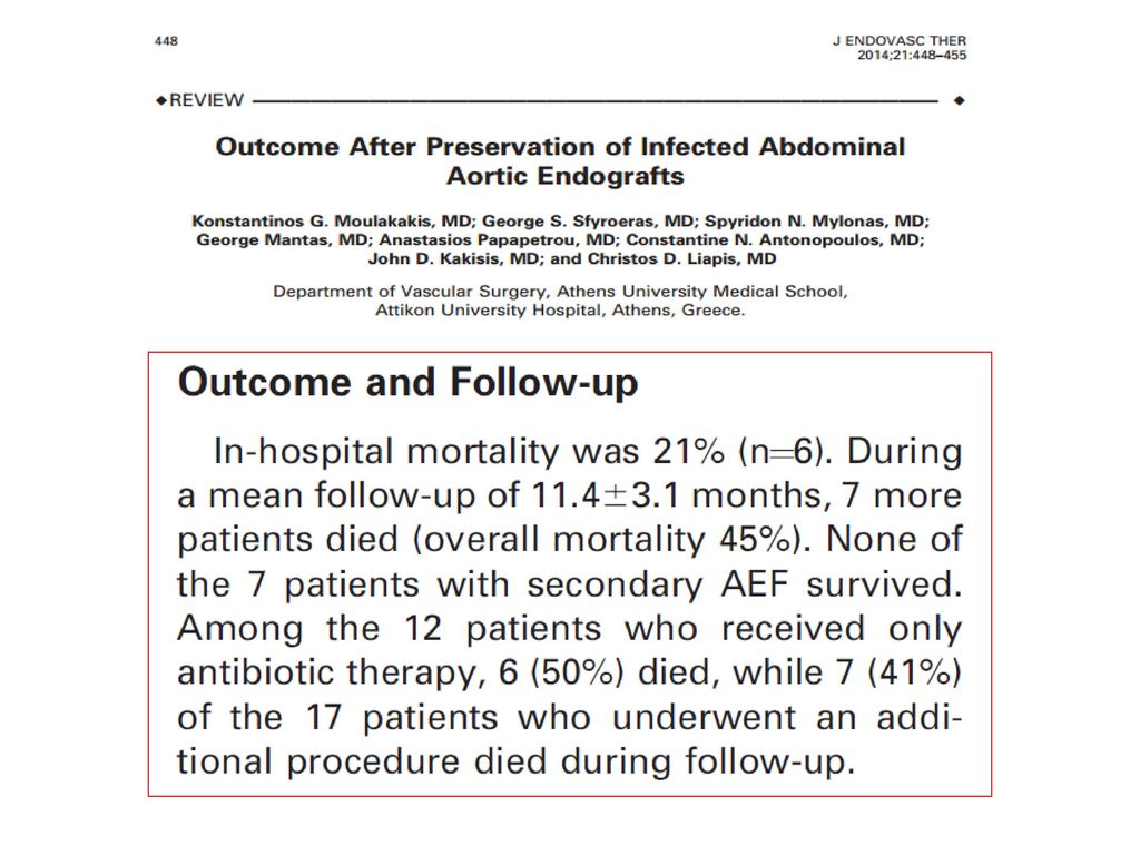
Broad spectrum antibiotics should administered as soon as a mycotic aneurysm is suspected Prolonged postoperative antibiotic therapy Additional procedures such as surgical debridement and percutaneous drainage are important adjuncts in eliminating the source of infection. 1. Moulakakis KG, Mylonas SN, Antonopoulos CN, Kakisis JD, Sfyroeras GS, Mantas G, Liapis CD. Comparison of treatment strategies for thoracic endograft infection. J Vasc Surg. 2014 2. Moulakakis KG, Sfyroeras GS, Mylonas SN, Mantas G, Papapetrou A, Antonopoulos CN, Kakisis JD, Liapis CD. Outcome after preservation of infected abdominal aortic endografts. J Endovasc Ther. 2014

22
Haemodynamic Instability, ↓ HgB
CASE 1 4,8 cm 83 y Fever up to 38.5 °C Leukocytosis: CRP: 136 Haemodynamic Instability, ↓ HgB Contained Rupture After 2 Days

23
After 24 Hours septic shock
EVAR Cook Zenith After 24 Hours septic shock Open laparotomy, drainage of the abscess, debridement around the endograft and irrigation with Garamycin

24
Abscess cultures St.Aureus
Discharged Cloxacillin Sodium and Rifampycin 3 month follow-up Moulakakis KG, Sfyroeras GS, Kakisis JD, Papapetrou A, Antonopoulos CN, Mantas G, Brountzos EN, Liapis CD. Endograft infection and treatment with preservation of the endograft: early results in 3 cases. Ann Vasc Surg. 2014

25
CASE 2 After 3 Days Fever up to 39 ° C Leukocytosis ↑ CRP
Thoracic pain Blood cultures (-) After 3 Days
 After 3 Days.")
26
TAA Diameter Increased
TEVAR VALIANT, MEDTRONIC

27
After 3 Months Fever up to 39 ° C Leukocytosis ↑ CRP
Esophagus stenting
28
Esophagectomy and Gastric pull
Discharged Cloxacillin Sodium and Rifampycin

29
Endograft infection Graft infection after endovascular aneurysm repair (EVAR or TEVAR) is an underrecognized and underreported event. The incidence of aorto-iliac stent-graft infection ranges from 0.4% to 0.7% (1). Although rare, it may have devastating consequences. Mortality rates range from 25% to 100% (0.6% to 3% for open aortic graft infection 2,3) Setacci C. et al. Management of abdominal endograft infection. J Cardiovasc Surg. 2010 T.W. Swain, et al. Management of infected aortic prosthetic grafts. Vasc Endovascular Surg. 2004 S. O’Connor, et al. A systematic review and meta-analysis of treatments for aortic graft infection. JVS.2006
. Although rare, it may have devastating consequences. Mortality rates range from 25% to 100% (0.6% to 3% for open aortic graft infection 2,3) Setacci C. et al. Management of abdominal endograft infection. J Cardiovasc Surg T.W. Swain, et al. Management of infected aortic prosthetic grafts. Vasc Endovascular Surg S. O’Connor, et al. A systematic review and meta-analysis of treatments for aortic graft infection. JVS")
30
Pathogenesis Stent Graft Related
Bacterial inoculation during endovascular procedure Pre-existing -mycotic aneurysm or inflammatory aneurysm-, could result in intestinal necrosis and fistula formation Remote source of sepsis (eg, endocarditis, pneumonia, urinary tract infection) Cancer or immunodeficiency Repeated secondary procedures Stent migration Erosion of the aorta and the duodenum by embolization coils Fabric rupture ?? Erosion of the aorta by the hooks and barbs Endoleak and endotension may lead to aorto-enteric fistula formation ?? Stent Graft Related Setacci C. et al. Management of abdominal endograft infection. J Cardiovasc Surg. 2010
 Cancer or immunodeficiency. Repeated secondary procedures. Stent migration. Erosion of the aorta and the duodenum by embolization coils. Fabric rupture Erosion of the aorta by the hooks and barbs. Endoleak and endotension may lead to aorto-enteric fistula formation Stent Graft Related. Setacci C. et al. Management of abdominal endograft infection. J Cardiovasc Surg")
31
Clinical Presentation
Aortic Endograft Infection Thoracic Endograft Infection Low grade infection Systemic Sepsis Aortoenteric Fistula (41%) Abdominal or back Pain Abscess (psoas) Pseudonaurysm Urinary tract infection Low grade infection Systemic Sepsis Fistula (aortoesophageal or broncial) (38%) Chest Pain Abscess (periaortic) Pseudonaurysm Pneumonia, mediastinitis 52%% 50%% Numan F. et al. Management of endograft infections. J Cardiovasc Surg. 2011
 Abdominal or back Pain. Abscess (psoas) Pseudonaurysm. Urinary tract infection. Low grade infection. Systemic Sepsis. Fistula (aortoesophageal or broncial) (38%) Chest Pain. Abscess (periaortic) Pseudonaurysm. Pneumonia, mediastinitis. 52%% 50%% Numan F. et al. Management of endograft infections. J Cardiovasc Surg")
32
Εμπλεκόμενοι Μικροοργανισμοί
20-83% αναγνωρίζεται και ταυτοποιείται ο υπεύθυνος μικροβιακός παράγοντας St.Aureus 22% Streptococcus sp. 11% Multiple pathogens 21% Candida Ablicans , Mycetes 6% E.Coli Enterococci Pseudomonas, Serratia, Klebsiella, Ent.Cloacae ↑f Numan F. et al. Management of endograft infections. J Cardiovasc Surg Setacci C. et al. Management of abdominal endograft infection. J Cardiovasc Surg. 2010

33
Διάγνωση μολυσμένου Μοσχεύματος
βαθμός υποψίας Καλλιέργειες Ενδοσκοπικός Ελεγχος (AEF) CT / MR PET CT Πυρηνικός Ελεγχος ,Σπινθηρογράγημα
 CT / MR. PET CT. Πυρηνικός Ελεγχος ,Σπινθηρογράγημα.")
34
Management of Infected Endograft
Depends on : Patient’s clinical status Co-morbidities Presence of preoperative sepsis Microorganisms involved

35
1. ΑΝΟΙΚΤΗ ΧΕΙΡΟΥΡΓΙΚΗ ΑΝΤΙΜΕΤΩΠΙΣΗ
1. ΑΝΟΙΚΤΗ ΧΕΙΡΟΥΡΓΙΚΗ ΑΝΤΙΜΕΤΩΠΙΣΗ

36
Management of Infected Endograft
Graft Excision is the GOLD STANDARD Graft Excision & Extra-anatomic bypass Neo-aortoiliac System Procedure In Situ Aortic Graft Replacement (Homograft, Silver Graft) High mortality and morbidity rates, especially when undertaken in unstable, septic patients with severe comorbidities Variable results on patency and reinfection rates Fiorani P, et al. Endovascular graft infection: preliminary results of an international enquiry. JEVT 2003
 High mortality and morbidity rates, especially when undertaken in unstable, septic patients with severe comorbidities. Variable results on patency and reinfection rates. Fiorani P, et al. Endovascular graft infection: preliminary results of an international enquiry. JEVT")
43
Technique of Aortic stent-graft explantation
JEVT 2010 Technique of Aortic stent-graft explantation Factors that may influence the feasibility of aortic stent-graft explantation The fixation system (hooks or barbs) the associated periaortic inflammatory reaction and endograft incorporation the presence of any additional grafts, cuffs, or coils placed as secondary interventions
 the associated periaortic inflammatory reaction and endograft incorporation. the presence of any additional grafts, cuffs, or coils placed as secondary interventions.")
44
2. ΠΑΡΟΧΕΤΕΥΣΗ, ΧΕΙΡΟΥΡΓΙΚΟΣ ΚΑΘΑΡΙΣΜΟΣ, ΔΙΑΤΗΡΗΣΗ ΤΟΥ ΜΟΣΧΕΥΜΑΤΟΣ
2. ΠΑΡΟΧΕΤΕΥΣΗ, ΧΕΙΡΟΥΡΓΙΚΟΣ ΚΑΘΑΡΙΣΜΟΣ, ΔΙΑΤΗΡΗΣΗ ΤΟΥ ΜΟΣΧΕΥΜΑΤΟΣ

45
Management of Infected Endograft in High Risk patients for open repair
Surgical or CT-guided percutaneous placement of drains into the aneurismal sac abscess contiguous to the graft, in conjunction with irrigation of the perigraft area followed by appropriate antibiotic therapy Promising results in patients without signs of severe sepsis Pryluck DS et al. Percutaneous drainage of aortic aneurysm sac abscesses following endovascular aneurysm repair.Vasc Endovascular Surg.2010 Deshmukh H. et al. Percutaneous management of complications (aortoenteric fistula and sac abscess) following bypass surgery for abdominal aortic aneurysm.Cardiovasc Intervent Radiol. 2007 S.J. Hulin* and G.E. Morris .Eur J Vasc Endovasc Surg .2007
 following bypass surgery for abdominal aortic aneurysm.Cardiovasc Intervent Radiol S.J. Hulin* and G.E. Morris .Eur J Vasc Endovasc Surg")
46
Management of Infected Endograft in High Risk patients for open repair
CT-guided percutaneous drainage followed by appropriate antibiotic therapy Pryluck DS et al. Percutaneous drainage of aortic aneurysm sac abscesses following endovascular aneurysm repair.Vasc Endovascular Surg.2010 Deshmukh H. et al. Percutaneous management of complications (aortoenteric fistula and sac abscess) following bypass surgery for abdominal aortic aneurysm.Cardiovasc Intervent Radiol. 2007 S.J. Hulin* and G.E. Morris .Eur J Vasc Endovasc Surg .2007
 following bypass surgery for abdominal aortic aneurysm.Cardiovasc Intervent Radiol S.J. Hulin* and G.E. Morris .Eur J Vasc Endovasc Surg")
47
Symptomatic 8.1 cm pararenal abdominal aortic aneurysm
CASE 1 A 63-year old man, smoker Hostile abdomen Previous MI AF under oral anticoagulants COPD Severe obesity (BMI: 36.6) EVAR 1 month CT type-Ia endoleak multiple coils were deployed followed by biological glue infusion resulting in successful type Ia endoleak treatment….BUT
 EVAR. 1 month CT type-Ia endoleak. multiple coils were deployed followed by biological glue infusion resulting in successful type Ia endoleak treatment….BUT.")
48
Eight months later…. Endograft Infection Fever up to 39,4 C
Lower back Pain Leucocytosis Increased CRP CTA Presence of air in the aneurysm sac cavity Blood Cultures: E.Coli and Ent. Faecalis Endograft Infection

49
Mini-Laparotomy : Sigmoid detached from the inflammatory mass, omentoplasty.
Percutaneous continuous drainage of aortic aneurysm sac abscess for 15 days CT- guided percutaneous continuous drainage followed by Vancomycin intrasac administration for 15 days Oral administration of moxifloxacin ( 400 mg daily dose x 30d)
50
Follow-up at 18 months CTA : No presence of air in the sac cavity
Decrease of aneurysm sac diameter Patient remains asymptomatic, afebrile. WBC : 5.300, CRP:9

51
Management of Infected Endograft in High Risk patients for open repair
63-year old man Infection 3 years after EVAR. Patient presented unstable, with sepsis and massive bleeding due to AEF CASE 2 Excision of the eroded part of the duodenum or the bowel and interposition of the omentum, without further aortic reconstruction, followed by antibiotic therapy.
52
interposition of the omentum, without further aortic reconstruction
Patient died on 3rd postoperative day due to MOF
53
Πρόγνωση Θνητότητας λόγω Μόλυνσης του Ενδομοσχεύματος
Fungal or gram negative species Presentation with severe sepsis AEF Bleeding requiring massive transfusion Advanced ASA physical status Age > 65 Renal insufficiency Stableford J. Endograft Infection after EVAR. October 2009

54
Ανασκόπηση της Βιβλιογραφίας
129 reported cases (36 post TEVAR, 93 post EVAR) Range of endograft infection 0.2-3% Mean Time to presentation 15.4 m (1-96 m) 33% Early < 4months % > 4 months Numan F. et al. Management of endograft infections. J Cardiovasc Surg Setacci C. et al. Management of abdominal endograft infection. J Cardiovasc Surg. 2010 Cernohorsky P.,JVS 2011
 Range of endograft infection 0.2-3% Mean Time to presentation 15.4 m (1-96 m) 33% Early < 4months - 67% > 4 months. Numan F. et al. Management of endograft infections. J Cardiovasc Surg Setacci C. et al. Management of abdominal endograft infection. J Cardiovasc Surg Cernohorsky P.,JVS")
55
Ανασκόπηση της Βιβλιογραφίας (Σειρές ≥ 3pts)
Author Pts Type of Endograft Procedure Schlensak C., JVS 2001 5 Stentor 2, Vanguard 3 EVAR Eggebrecht H., JEVT 2004 3 N.D. TEVAR Sharif MA, JVS 2007 6 Zenith 4, Talent 2 Brown KE.,JVS 2008 TAG 5, Cuff 1 Girdauskas E,J.Thor.Card.Surg. 2008 4 Sarantzis N., JEVT 2008 Ebdofit 3, Anaconda 1, Powerlink 1 Heyer KS, JVIR 2009 10 TAG 4, Excluder 3, Zenith 2, Ancure 1 5 EVAR 5 TEVAR Kelso RL, JVS 2009 Excluder 2, AneuRx 1, Ancure 1 Chiesa R.,J.Card.Surg.2010 7 Zenith 3, Endofit 1, TAG 1, Relay 1, N.D. 1 Cernohorsky P.,JVS 2011 12 Zenith 3, Talent 9 2 TEVAR/ 10 EVAR

56
3/4 pts treated conservatively
Author Pts Management Mortality Schlensak C. 5 Stent Removal and Extra-an. BP (5) Not Described Eggebrecht H. 3 Conservative (3) 100% Sharif MA 6 Stent Removal and Extra-an. BP (3) Conservative (2) None (Early Death) (1) 50% Brown KE. Stent Removal and Extra-an. BP (1) Conservative (1) Unknown (4) 66.6% Girdauskas E 4 Stent Removal and reconstruction (4) 25% Sarantzis N. None (Early Death) (2) 60% Heyer KS 10 Conservative (4) Stent Removal and reconstruction (3) 30% 3/4 pts treated conservatively Kelso RL Stent Removal and Extra-an. BP (2) Stent Removal and aortobifemoral (1) Inraoperative Death (1) 1 Death 3 Lost in FU Chiesa R. 7 Stent Removal and reconstruction (2) Conservative (1) None (periop. Death) (3) 71% Cernohorsky P. 12 Surgical Repair (6) Conservative (6)
 Not Described. Eggebrecht H. 3. Conservative (3) 100% Sharif MA. 6. Stent Removal and Extra-an. BP (3) Conservative (2) None (Early Death) (1) 50% Brown KE. Stent Removal and Extra-an. BP (1) Conservative (1) Unknown (4) 66.6% Girdauskas E. 4. Stent Removal and reconstruction (4) 25% Sarantzis N. None (Early Death) (2) 60% Heyer KS. 10. Conservative (4) Stent Removal and reconstruction (3) 30% 3/4 pts treated conservatively. Kelso RL. Stent Removal and Extra-an. BP (2) Stent Removal and aortobifemoral (1) Inraoperative Death (1) 1 Death. 3 Lost in FU. Chiesa R. 7. Stent Removal and reconstruction (2) Conservative (1) None (periop. Death) (3) 71% Cernohorsky P. 12. Surgical Repair (6) Conservative (6)")
57
Review of the literature Clinical Outcomes in 102 pts
Overall Mortality 40.1% TEVAR 64.7%, EVAR 30.1% TEVAR EVAR Surgical Treat. Conservative Mortality (8/17) 47.1% (7/10) 70% 17/ % (4/6) 66.6% Numan F. et al. Management of endograft infections. J Cardiovasc Surg
 47.1% (7/10) 70% 17/ % (4/6) 66.6% Numan F. et al. Management of endograft infections. J Cardiovasc Surg")
58
Review of the literature Clinical Outcomes in 102 pts
EVAR- Surgical Repair Overall Mortality (17/60) 28.3% aorta ligation and extra anatomic BP (42) - Mortality (13/42) : 30.9% In situ reconstruction (18) - Mortality ( 4 /18) : 22.2% TEVAR - Surgical Repair Thor. aorta ligation and extra-anatom. BP (5) In situ reconstruction (9) Esophageal or bronchial repair (3) Overall Mortality (8/17) 47.1%
 28.3% aorta ligation and extra anatomic BP (42) - Mortality (13/42) : 30.9% In situ reconstruction (18) - Mortality ( 4 /18) : 22.2% TEVAR - Surgical Repair. Thor. aorta ligation and extra-anatom. BP (5) In situ reconstruction (9) Esophageal or bronchial repair (3) Overall Mortality (8/17) 47.1%")
59
Μόλυνση Ενδομοσχεύματπος μετά από EVAR

60
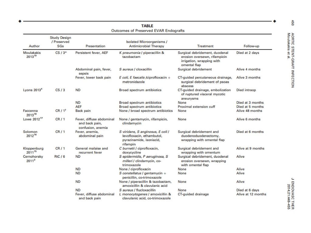
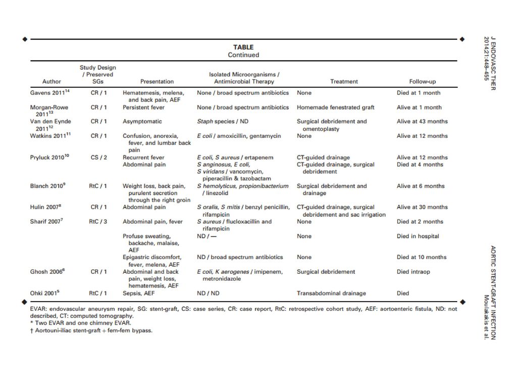
N of patients treated with endograft preservation
Author Study design Type of procedure N of patients treated with endograft preservation Moulakakis et al, case series 2EVAR, 1ch-EVAR 3 Lyons et al, 20134 EVAR Faccenna et al, case report AUI + fem-fem 1 Lowe et al, Solomon et al, Kloppenburg et al,201116 Cernohorsky et al, 20113 Retr. cohort study 6 Gavens et al, EVAR/TEVAR Morgan-Rowe et al, FEVAR Van den Eynde et al, Watkins et al, Pryluck et al, 2 Blanch et al, Saleem et al, 20089 Hulin et al,20078 Sharif et al, 20077 Ghosh et al, 20066 Ohki et al, 20015 Total 30

61
Μόλυνση Ενδομοσχεύματπος μετά από EVAR
Number of patients 30 Gender (%male) 94 Age (years, mean ± SE) 72.8 ±8.4 Setting of the EVAR procedure ● Elective (%) ● Emergent (%) 82 18 Reintervention (%) 13 Time to infection (mean, days) 360±81
 94. Age (years, mean ± SE) 72.8 ±8.4. Setting of the EVAR procedure. ● Elective (%) ● Emergent (%) Reintervention (%) 13. Time to infection (mean, days) 360±81.")
65
Infected Endograft as “bridging “ procedure after Aortoenteric Fistula
67
The Graft Must Come Out: Is there alternative option ?
High mortality and morbidity rates, especially when undertaken in unstable, septic patients with severe comorbidities The Graft Must Come Out: What If It Can't? Is there alternative option ?

68
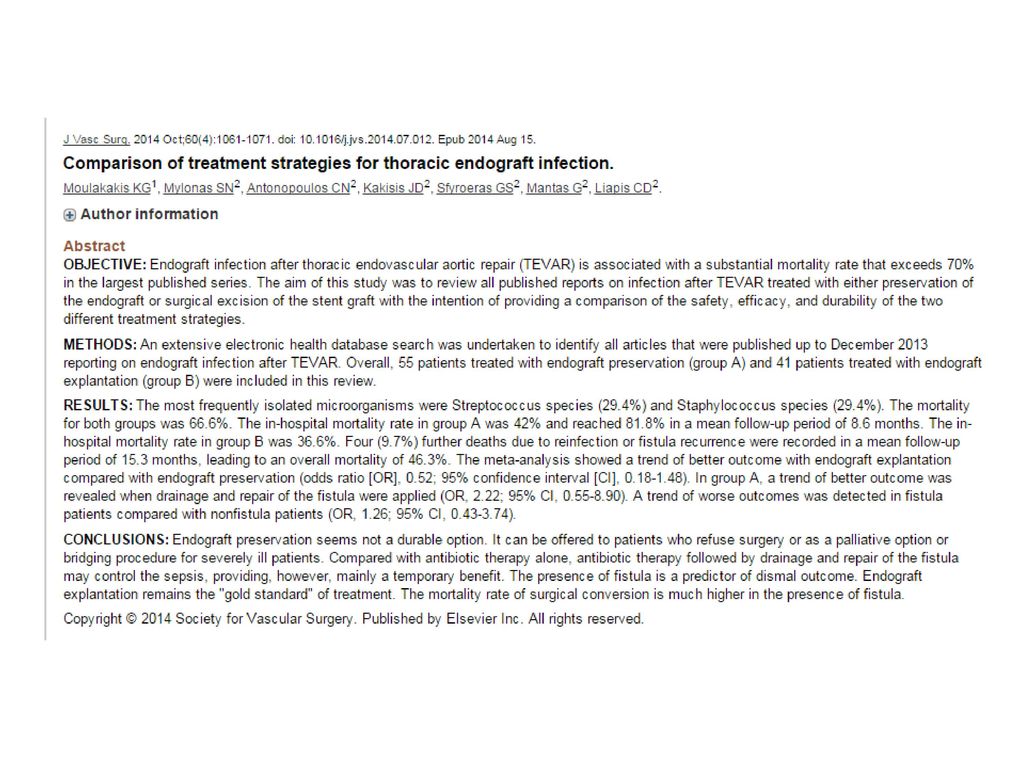
Comparison of treatment strategies for thoracic endograft infection
END-POINTS 1. Endograft explantation vs endograft preservation; 2. Additional treatment vs. none in patients treated with endograft preservation 3. Presence of fistula vs absence of fistula. Review Metanalysis Group A: 55 pts treated with endograft preservation Group B: 41 pts treated with endograft explantation (group B) Moulakakis KG, Mylonas SN, Antonopoulos CN, Kakisis JD, Sfyroeras GS, Mantas G, Liapis CD. Comparison of treatment strategies for thoracic endograft infection. J Vasc Surg. 2014
 Moulakakis KG, Mylonas SN, Antonopoulos CN, Kakisis JD, Sfyroeras GS, Mantas G, Liapis CD. Comparison of treatment strategies for thoracic endograft infection. J Vasc Surg")
69
Μόλυνση Ενδομοσχεύματος - Συμπεράσματα
5. Στην μόλυνση μετα από TEVAR, η διατήρηση του μοσχεύματος έχει εξαιρετικά απογοητευτικά αποτελέσματα 6. Όταν συνυπάρχει επικοινωνία η ενδαγγειακή αποκατάσταση συνίσταται για τον έλεγχο της αιμορραγίας, την βελτίωση της γενικής κατάστασης του ασθενή και σαν γέφυρα για μελλοντική εξαίρεση του μοσχεύματος.

Παρόμοιες παρουσιάσεις


 : ΑΝΟΙΧΤΩΝ Vs ΚΛΕΙΣΤΩΝ ΚΥΨΕΛΩΝ>")


. ΣΥΓΚΟΠΤΙΚΗ ΚΡΙΣΗ Αιφνίδια και σύντομη απώλεια συνείδησης που προκαλείται από ανεπαρκή εγκεφαλική αιμάτωση λόγω μειωμένης.>")


 ωφελέειν ή μη βλάπτειν ωφελέειν = θεραπευτική παρέμβαση μη βλάπτειν = ασφάλεια ασθενών.>")


