Κατέβασμα παρουσίασης
Η παρουσίαση φορτώνεται. Παρακαλείστε να περιμένετε

1
Διαχείριση αεραγωγού – CPR
Πέτρος Κοπτερίδης 2η Κλινική Εντατικής Θεραπείας - «Αττικό» Νοσοκομείο Νοέμβριος 2011

2
Διαχείριση αεραγωγού

3
Σκοπός της διαχείρισης αεραγωγού
Εξασφάλιση ανεμπόδιστης ανταλλαγής αερίων Αποφυγή πλήρωσης αεραγωγών με αίμα, τροφή κ.λ.π. Η ΑΠΟΦΡΑΞΗ ΑΕΡΑΓΩΓΟΥ ΑΠΑΙΤΕΙ ΕΠΕΙΓΟΥΣΑ ΠΑΡΕΜΒΑΣΗ Συνήθως έχετε < 5 min στην διάθεσή σας (Γιατί?) Even endotracheal intubation does not stop MICROSCOPIC aspiration (keep intubated patients sitting up at degrees)
 Even endotracheal intubation does not stop MICROSCOPIC aspiration (keep intubated patients sitting up at degrees)")
4
Many techniques to achieve airway patency are available
Many techniques to achieve airway patency are available. Choice of airway technique depends on 4 main issues: Good personal skill level with the technique chosen, even if an alternative technique might have theoretical advantages Need for definitive airway control Available equipment (always check your working environment to ensure you are familiar with airway equipment) Availability of skilled assistance from someone able to use or supervise the use of alternative techniques
 Availability of skilled assistance from someone able to use or supervise the use of alternative techniques.")
5
Προετοιμασία Essential equipment must be prepared and checked to make sure it is working. Faulty equipment is associated with adverse events and adverse events potentially become catastrophic when worsened by missing or faulty equipment: Some examples: Oxygen - Suction - Monitoring Equipment - Drugs (including iv access)
6
Προετοιμασία Μιλήστε στον ασθενή / Εξηγήστε του
Φωνάξτε βοήθεια έγκαιρα In awake patients, explanation of what you are about to do is essential to relieve anxiety. If you lack experience and there is any possibility of summoning experienced help then help should be summoned immediately. Any kind of help is useful if you run into difficulty during intubation. IT CANNOT BE OVERSTRESSED - DO NOT HESITATE TO CALL FOR HELP IF YOU THINK IT MAY BE NECESSARY

7
Βασική διαχείριση αεραγωγού
Head tilt - Chin lift Airway management can be divided into BASIC & ADVANCED MANAGEMENT Basic airway maneuvers Relieving obstruction by the head tilt is easy and effective. A hand firmly placed on the forehead tilts the head backward on the atlanto-occipital joint. The chin lift is completed by placing the fingers of the one hand under the bony part of the lower jaw and lifting the chin forward. Note: The tongue and posterior pharynx is lifted away from the posterior pharynx

8
Τriple airway maneuver
The head tilt, jaw thrust, mouth open (triple airway maneuver) is used when other methods have failed to open the airway. The head is tilted back in extension and the fingers of both hands grasp the ramus of the mandible which is displaced forward and upward. Both thumbs are then used to open the lower lips. BEWARE OF PATIENTS WITH CERVICAL SPINE INJURIES
 is used when other methods have failed to open the airway. The head is tilted back in extension and the fingers of both hands grasp the ramus of the mandible which is displaced forward and upward. Both thumbs are then used to open the lower lips. BEWARE OF PATIENTS WITH CERVICAL SPINE INJURIES.")
9
Στοματο-φαρυγγικοί αεραγωγοί
Oropharyngeal airways may be useful to prevent soft tissues from obstructing the airway in the unconscious patient at the levels of the soft palate, epiglottis and base of tongue. An oro-pharyngeal airway may establish an adequate airway for spontaneous or bag-mask ventilation when proper head positioning is insufficient. It is inserted with the concavity facing the palate and then rotated 1800 into the proper position as it is advanced. Complications: Gagging, laryngospasm, or vomiting. Mucosal trauma, worsening the obstruction by pressing the epiglottis against the laryngeal outlet or displacing the tongue more posteriorly.

10
Ρινο-φαρυγγικοί αεραγωγοί
A nasopharyngeal airway is a soft rubber or plastic tube inserted into the nostril and advanced horizontally along the floor of the nose into the posterior pharynx. Functions similarly to the oro-pharyngeal airway, but is often better tolerated by semi-conscious patients. Complications: Epistaxis, aspiration, laryngospasm and esophageal placement Insert Horizontally

11
15L/min Once obstruction has been resolved it may be possible to allow the patient to breathe spontaneously (the left lateral position is recommended). Usually some form of ventilatory assistance is required. In this setting a manual bag valve resuscitator is usually used. Requires time and experience to master technique - skill station. Most resuscitators are self-inflating with a separate reservoir bag in series that ensures a consistent oxygen concentration. Note valve arrangement - must know how the valves work (skill station) and test to make sure the assembly works before use. Wall flow must be adequate to keep reservoir bag inflated (usually 15 l/min). Self inflating part and reservoir. The addition of positive end-expiratory pressure (PEEP) valves may improve arterial oxygenation and help to overcome airway obstruction due to laryngospasm. Transparent masks are recommended
. Usually some form of ventilatory assistance is required. In this setting a manual bag valve resuscitator is usually used. Requires time and experience to master technique - skill station. Most resuscitators are self-inflating with a separate reservoir bag in series that ensures a consistent oxygen concentration. Note valve arrangement - must know how the valves work (skill station) and test to make sure the assembly works before use. Wall flow must be adequate to keep reservoir bag inflated (usually 15 l/min). Self inflating part and reservoir. The addition of positive end-expiratory pressure (PEEP) valves may improve arterial oxygenation and help to overcome airway obstruction due to laryngospasm. Transparent masks are recommended.")
12
Tidal volume: 300-500 ml + RR: 10-16/min
Tips and pitfalls Gastric insufflation is common and increases the risk of vomiting and aspiration. Severe intra-abdominal distension may cause cardiovascular compromise. Carefully applied cricoid pressure may prevent gastric gas insufflation Over-distention of the lungs. Tidal volumes of L can be delivered. Rapid delivery of high tidal volumes can easily lead to gas being retained in the lung. Over-distention and high intra-thoracic pressures may cause profound hemodynamic instability Tips: Concentrate on providing tidal volumes of only about ml at respiratory rates of 10-16/min In the emergency situation with a full stomach, mask ventilation with an assistant providing cricoid pressure may be necessary until the airway can be secured

13
χρειάζονται 2 ανανήπτες
Σε περιπτώσεις που δεν γίνεται καλή επαφή της μάσκας με το πρόσωπο (π.χ. ασθενείς με μούσι ή χωρίς οδοντοστοιχία) ανεπαρκής αερισμός χρειάζονται 2 ανανήπτες
 ανεπαρκής αερισμός χρειάζονται 2 ανανήπτες.")
14
Τι μπορεί να οδηγήσει σε ενδοτραχειακή διασωλήνωση για έλεγχο του αεραγωγού
Ο ασθενής δε μπορεί μόνος του να διατηρήσει βατό τον αεραγωγό του Απαιτείται υψηλή PEEP ή παράλυση Ανάγκη για συνεχείς αναρροφήσεις It is now possible to provide mechanical ventilation to patients with respiratory failure with non-invasive methods. Respiratory failure by itself is therefore no longer an absolute indication for endotracheal intubation. Examples : Airway obstruction Unconsciousness (own soft tissues cause obstruction etc) Airway obstruction (swelling from anaphylaxis, external compression from hematoma etc) Severe metabolic acidosis (exhaustion expected) Mechanical ventilation with high PEEP or paralysis ARDS - massive aspiration Patient requires airway suction Muscle weakness, myasthenia gravis, large particle aspiration etc
 Airway obstruction (swelling from anaphylaxis, external compression from hematoma etc) Severe metabolic acidosis (exhaustion expected) Mechanical ventilation with high PEEP or paralysis. ARDS - massive aspiration. Patient requires airway suction. Muscle weakness, myasthenia gravis, large particle aspiration etc.")
15
During intubation the patient must be adequately monitored
ECG BP SaO2 ETCO2 Monitoring During intubation the patient must be adequately monitored Basic monitoring includes: Oximetry - ECG rhythm monitor - Capnography - Blood pressure (invasive or non-invasive) Clinical: Chest movement/breath sounds - Cyanosis - Heart rate
 Clinical: Chest movement/breath sounds - Cyanosis - Heart rate.")
16
Ενδοτραχειακοί σωλήνες
Κύκλωμα Λαρυγγοσκόπια Αεραγωγοί Φάρμακα ΠΡΟΕΤΟΙΜΑΣΙΑ Essential intubation equipment must be prepared and checked (tricks to remember ‘x plastics/y metals/suction etc) Standard laryngoscope and blades (Macintosh sizes 2, 3 and 4) Bougie & stylet Syringe for cuff inflation Magill forceps, large artery forceps Oral airways Endotracheal tubes of various sizes Catheter mount (device to connect circuit to endotracheal tube) Drugs Suction Λαβίδα Ενδοτραχειακοί σωλήνες Αναρρόφηση
 Standard laryngoscope and blades (Macintosh sizes 2, 3 and 4) Bougie & stylet. Syringe for cuff inflation. Magill forceps, large artery forceps. Oral airways. Endotracheal tubes of various sizes. Catheter mount (device to connect circuit to endotracheal tube) Drugs. Suction. Λαβίδα. Ενδοτραχειακοί σωλήνες. Αναρρόφηση.")
17
Intubation results in bypassing of all infectious defenses of the upper airways - keep the procedure as sterile or at least as clean as possible

18
Προ-οξυγόνωση Head position is critical to success of the procedure. The neck flexed (best achieved by elevating the occiput about 7-10 cm with a form pillow or wedge) and head extended Notes Pre-oxygenate with 100% oxygen for 2-5 min / In-line stabilization for patients with cervical spine injury or instability 7-10 cm
 and head extended. Notes. Pre-oxygenate with 100% oxygen for 2-5 min / In-line stabilization for patients with cervical spine injury or instability cm.")
19
Καταστολή και μυοχάλαση
Μόνο αν είναι απαραίτητο Πρέπει να είστε σίγουροι ότι μπορείτε να «χειριστείτε» τον αεραγωγό Προσοχή στις παρενέργειες της σουκινυλοχολίνης Ροκουρόνιο: εναλλακτικό μυοχαλαρωτικό μακράς δράσης Intubating conditions are variable. You need to judge whether drugs are required to improve intubating conditions. They must always be prepared before-hand (with a working IV) - even if you do not anticipate using them
 - even if you do not anticipate using them.")
20
Λαρυγγοσκόπιο Note horizontal and vertical parts of blade. Vertical part allows the operator to move the tongue to the left. The horizontal part allows the tongue/epiglottis to be elevated at the appropriate time. Note the light is on and bright

21
Λάρυγγας Important landmarks that you are aiming to see during direct laryngoscopy: Epiglottis - Vocal cords - Arytenoid cartilages

22
Rapid Sequence Intubation
Προ-οξυγόνωση Πίεση στον κρικοειδή χόνδρο Bolus φαρμάκων Άμεση διασωλήνωση Άρση πίεσης στον κρικοειδή χόνδρο όταν επιβεβαιωθεί η σωστή θέση του ενδοτραχειακού σωλήνα Most ICU patients are at risk of aspiration of gastric contents during intubation and therefore rapid sequence induction and intubation is required

23
Πίεση στον κρικοειδή χόνδρο
Note the anatomical position of the cricoid ring. Candidates to find it (below) caudal to the crico-thyroid membrane on themselves. Three fingers - outside to steady and middle to provide pressure. Note esophageal occlusion
 caudal to the crico-thyroid membrane on themselves. Three fingers - outside to steady and middle to provide pressure. Note esophageal occlusion.")
24
Τεχνική της διασωλήνωσης
Note position of epiglottis at base of tongue. Note laryngoscope blade is in front of the epiglottis and not covering it. The final position of the head and the laryngoscope blade allows a straight line for vision and insertion of the endotracheal tube.

25
Τεχνική της διασωλήνωσης
With good head position, advance the laryngoscope blade (in your left hand) gently along the right hand side of the tongue until the tip of the epiglottis is seen
 gently along the right hand side of the tongue until the tip of the epiglottis is seen.")
26
Τεχνική της διασωλήνωσης
Advance the laryngoscope blade to reveal the epiglottis

27
Τεχνική της διασωλήνωσης
The blade tip is placed in the groove between the base of the tongue and the epiglottis
28
Τεχνική της διασωλήνωσης
Once in position - lift the laryngoscope in the direction of the handle. This will reveal the cords behind the epiglottis

29
Τεχνική της διασωλήνωσης
Note the anatomical positions again

30
Τεχνική της διασωλήνωσης
The entire tongue is lifted in the direction of the laryngoscope handle and the cords are revealed behind the epiglottis. Extra points: Describe the BURP maneuver

31
The BURP maneuver Backward-Upward-Rightward Pressure of the larynx

32
Τεχνική της διασωλήνωσης
The endotracheal tube is inserted from the right side with the right hand - Confirm position by direct visualization of the vocal cords and passage of the tube

33
Επιβεβαίωση θέσης του τραχειοσωλήνα
It is still possible to make a mistake and place the tube in the esophagus Measurement of expired CO2 by capnography is an accurate way to confirm tube position in the trachea. Carbon dioxide is only produced in the lungs and therefore only expired gas can contain CO2 Note that in a pre-oxygenated patient SaO2 may remain high for some minutes, even after mistaken esophageal intubation Tips Capnography may produce false-positive results (detectable end-tidal CO2) with the first few breaths after inadvertent esophageal intubation (this is possible if gastric insufflation from prior mask ventilation has occurred) A false-negative (decreased or very low CO2 despite correct position) may occur with cardiac arrest and low cardiac output states
 with the first few breaths after inadvertent esophageal intubation (this is possible if gastric insufflation from prior mask ventilation has occurred) A false-negative (decreased or very low CO2 despite correct position) may occur with cardiac arrest and low cardiac output states.")
34
Επιβεβαίωση θέσης του τραχειοσωλήνα
Clinical signs such as auscultation of breath sounds over the chest and epigastrium, are useful, but less reliable Auscultation is also useful to detect a tube that is too deep and in the right main bronchus

35
Επιβεβαίωση θέσης του τραχειοσωλήνα
Most male patients require the endotracheal tube to be tied at about 23 cm from the tip. Most female patients require the endotracheal tube to be tied at about 21 cm from the tip. Tip Auscultation of breath sounds over the chest and epigastrium may be useful to detect a tube that is too deep and in a main bronchus Most commonly decreased breath sounds on the left side are heard, indicating a right main bronchus intubation

36
Secure endotracheal tube carefully with tape
37
Αποτυχημένη διασωλήνωση
1-5% Η επακόλουθη υποξαιμία μπορεί να έχει δραματικές συνέπειες Πρέπει να έχετε έτοιμο το σχέδιο Β

38
No anticipated intubation difficulty
Rapid sequence induction Succeed Direct laryngoscopy and intubation Fail Call for help Able to manually ventilate No Insert laryngeal mask Yes Ventilation Yes No Algorithms are designed to prepare decisions for you to help deal with difficult situations that can be anticipated. Only relevant pathways need to be considered for individual patients. This is a very simple algorithm for endotracheal intubation failure. More complex and comprehensive algorithms are available from various sources Reposition patient. One more D.L. attempt Fail Confirm tube position & ventilate patient Succeed Maintain ventilation wake up patient or advanced airway techniques* Surgical Airway * If experienced help available

39
Λαρυγγική μάσκα LMAs are useful to achieve non-definitive airway patency in many emergency situations. A LMA is prepared for insertion by deflating and smoothing out the cuffed rim to be wrinkle-free. The posterior surface is lubricated with water-soluble jelly. The patient is positioned in a similar position to that for endotracheal intubation - slight flexion of the neck and extension of the atlanto-occipital joint (“sniffing the morning air” position). The LMA is inserted with the tip of the cuff continuously applied to the hard palate and with the right index finger guiding the tube to the back of the tongue
. The LMA is inserted with the tip of the cuff continuously applied to the hard palate and with the right index finger guiding the tube to the back of the tongue.")
40
Λαρυγγική μάσκα Continue applying pressure until a firm resistance is encountered The cuff is then inflated with ml of air (adult sizes) Test for correct positioning (central positioning and unobstructed gas exchange, ETCO2) Attach to the breathing circuit
 Attach to the breathing circuit.")
41
Επείγων χειρουργικός αεραγωγός
Κρικοθυρεοειδοτομή Επείγων χειρουργικός αεραγωγός

42
Δύσκολος αεραγωγός Βραχύς λαιμός, παχύσαρκος ή μυώδης (θυρο-πωγωνική απόσταση < 6 cm) Περιορισμός κίνησης αυχένα - γνάθου Πρόπτωση οδόντων, μικρό στόμα, τοξοειδής επιμήκης υπερώα ή εισολκή κάτω γνάθου Απόφραξη στοματο-φάρυγγα και λάρυγγα Anticipating a difficult airway.

43
Obvious difficulty expected
Obvious difficulty expected. Fiberoptic intubation used with ENT surgeon on stand-by for emergency tracheostomy
44
Αναμενόμενος δύσκολος αεραγωγός
Φωνάξτε βοήθεια ΑΜΕΣΑ ενώ επιτελείτε βασικούς χειρισμούς αεραγωγού Υψηλή ροή χορηγούμενου οξυγόνου με AMBU Προετοιμαστείτε για «εξειδικευμένους» χειρισμούς αεραγωγού Αναμείνατε βοήθεια Αν ο ασθενής παρουσιάσει πλήρη απόφραξη αεραγωγού ή καρδιο-αναπνευστικό arrest , προσπαθήστε να απελευθερώσετε τον αεραγωγό με ό,τι τεχνική είστε πιο εξοικειωμένος

45
Περίληψη Εκπαίδευση Προσομοίωση (simulation) Εμπειρία Επανεκπαίδευση
 Εμπειρία Επανεκπαίδευση")
46
Cardio-pulmonary resuscitation Cardio-cerebral resuscitation
CPR Cardio-pulmonary resuscitation Cardio-cerebral resuscitation

47
Ασθενής που (πέφτει στο έδαφος και) χάνει τις αισθήσεις του
Φωνάξτε βοήθεια Απελευθερώστε τον αεραγωγό Απομακρύνετε από το στόμα ξένα σώματα, τροφή κ.λ.π.

48
Ασθενής που (πέφτει στο έδαφος και) χάνει τις αισθήσεις του
Ελέγξτε για αναπνοή Αγωνιώδεις αναπνευστικές προσπάθειες είναι σημείο ανακοπής Ελέγξτε για σφυγμό

49
ΟΧΙ σφυγμός - ΟΧΙ αναπνοή
ΒΟΗΘΕΙΑ

50
ΟΧΙ σφυγμός - ΟΧΙ αναπνοή
Ξεκινήστε θωρακικές συμπιέσεις Στο κατώτερο μισό του στέρνου / Συμπίεση 4-5 cm 100 /min 50% συμπίεση - 50% επαναφορά American Heart Association: 2 rescue αναπνοές υπό προϋποθέσεις Αερισμός 2 αναπνοές μετά από 30 θωρακικές συμπιέσεις 1 sec / αναπνοή “Φυσιολογική” έκπτυξη θώρακα

51
2 ανανήπτες 30:2

52
Άφιξη απινιδωτή ΕΛΕΓΞΤΕ ΤΟ ΡΥΘΜΟ Αυτοκόλλητα pads
Defibrillation paddles ΗΚΓ ηλεκτρόδια

54
Φορτίστε τον απινιδωτή
Συνεχίστε συμπιέσεις ενώ φορτίζεται ο απινιδωτής

55
DC shock 360 J (monophasic) 150-360 J (biphasic)
Leave ventilation bag connected to ETT. If patient is not intubated move it at least 1 m from patient’s chest during defibrillation

56
Ξαναρχίστε θωρακικές συμπιέσεις και αερισμό
Ξαναρχίστε θωρακικές συμπιέσεις και αερισμό ΜΗΝ ΕΛΕΓΞΕΤΕ ΤΟ ΣΦΥΓΜΟ ΣΥΝΕΧΙΣΤΕ ΓΙΑ 2 min

57
Εν μέσω θωρακικών συμπιέσεων και αερισμού
Αναγνωρίστε και θεραπεύστε αναστρέψιμες αιτίες ανακοπής

58
Δυνητικά αναστρέψιμες αιτίες ανακοπής: 4 Hs + Ts
Hypoxia Hypovolemia Hyperkalemia, hypokalemia, hypocalcemia, acidemia & other metabolic disorders Hypothermia Tension pneumothorax Tamponade Toxic substances Thromboembolism (Πνευμονική εμβολή - Έμφραγμα)
")
59
ΕΠΑΝΕΛΕΓΞΤΕ ΤΟ ΡΥΘΜΟ DC shock
360 J monophasic J biphasic

60
Ξαναρχίστε θωρακικές συμπιέσεις και αερισμό
Ξαναρχίστε θωρακικές συμπιέσεις και αερισμό ΜΗΝ ΕΛΕΓΞΕΤΕ ΤΟ ΣΦΥΓΜΟ ΣΥΝΕΧΙΣΤΕ ΓΙΑ 2 min

61
ΕΠΑΝΕΛΕΓΞΤΕ ΤΟ ΡΥΘΜΟ DC shock Αδρεναλίνη
Ξαναρχίστε θωρακικές συμπιέσεις και αερισμό για 2 min

62
Αδρεναλίνη 1 mg Αν επιμένει VF/VT μετά το 2ο shock
Ακολούθως: κάθε 3-5 min

63
Φλεβοκομβική ταχυκαρδία
ΕΠΑΝΕΛΕΓΞΤΕ ΤΟ ΡΥΘΜΟ Φλεβοκομβική ταχυκαρδία Ελέγξτε σφυγμό Υπάρχει σφυγμός Ελέγξτε πίεση 90/50 mm Hg

64
Αντιμετώπιση όσων επιβιώνουν της ανακοπής
Αντιμετώπιση όσων επιβιώνουν της ανακοπής Θεραπεύστε την αιτία Υποστηρίξτε τη λειτουργία των οργάνων Αντιμετωπίστε επιληπτικές κρίσεις Αποφύγετε την υπεργλυκαιμία Σκεφτείτε θεραπευτική υποθερμία

66
Αντενδείξεις θεραπευτικής υποθερμίας
Ενδείξεις θεραπευτικής υποθερμίας Εκτός νοσοκομείου VF ανακοπή Πιθανώς και: Άλλα είδη ανακοπής εκτός νοσοκομείου Ανακοπή εντός νοσοκομείου Αντενδείξεις θεραπευτικής υποθερμίας Ice packs Καρδιογενές shock Διαταραχές πήξης (η θρομβόλυση δεν θεωρείται αντένδειξη)
")
67
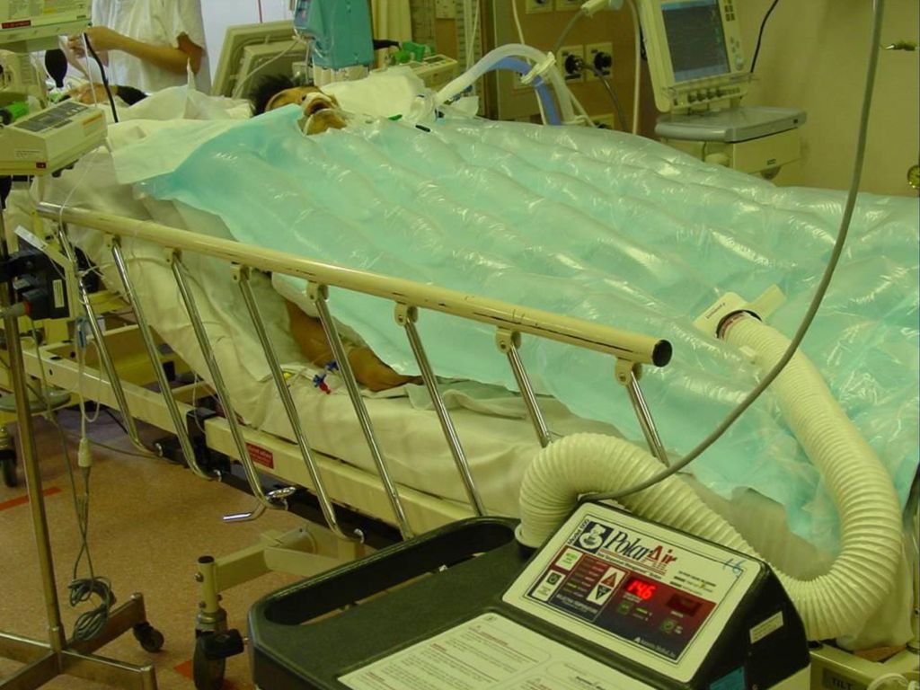
Μέθοδοι επίτευξης υποθερμίας
Εξωτερικές μέθοδοι: Κουβέρτες υποθερμίας – Ψυχρά επιθέματα Καταστολή & Παράλυση Συνεχές monitoring της θερμοκρασίας Στόχος 32-34C 4-6 ώρες μετά την ανακοπή Διατήρηση υποθερμίας για 24 ώρες Ice packs

68
Άλλα θέματα

69
Αμιοδαρόνη 300 mg bolus αν VF/VT επιμένει μετά από 3 shocks
Κάποιοι προτείνουν extra 150 mg bolus για ανθεκτική/υποτροπιάζουσα VF/VT Εν συνεχεία: Στάγδην έγχυση Give amiodarone if after the third shock the patient is still in VF

70
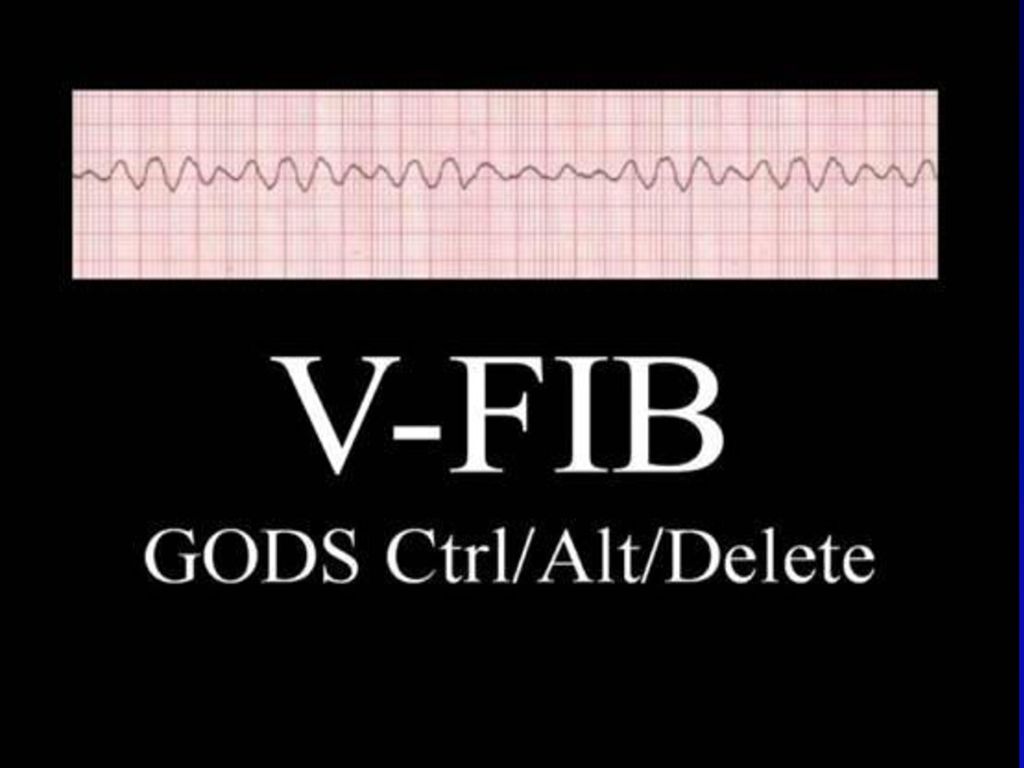
«Λεπτή» κοιλιακή μαρμαρυγή
ΔΔ: Ασυστολία ή Κοιλιακή μαρμαρυγή? UK Resuscitation Council Αν υπάρχει αμφιβολία, ΜΗΝ απινιδώσετε Αντιμετωπίστε σαν ασυστολία με αδρεναλίνη, συμπιέσεις και αερισμό AHA 2 min/5 κύκλοι CPR Απινιδώστε μια φορά

71
Διασωλήνωση κατά την ανακοπή
Αμφιλεγόμενη χρησιμότητα Αν επιχειρηθεί πριν την «επιστροφή» αυτόματης κυκλοφορίας: Μόνο από έμπειρους Συνέχιση συμπιέσεων κατά τη λαρυγγοσκόπηση Διακοπή συμπιέσεων μόνο για τοποθέτηση ενδοτραχειακού σωλήνα Όχι προσπάθεια > 30 sec

72
Πρακτικές συμβουλές (για αποφυγή διασποράς λοιμωδών παραγόντων κατά την διάρκεια της CPR)
Αρχίστε συμπιέσεις ενώ οι υπόλοιποι φορούν προστατευτικό εξοπλισμό: Face shield Fit -tested N95 mask Gown - Gloves

73
Πρακτικές συμβουλές Τεχνική αερισμού με 2 ανανήπτες Τοποθέτηση φίλτρου

74
Πρακτικές συμβουλές Προσοχή με τα μολυσμένα υλικά
Προσεκτικό πλύσιμο μετά τις προσπάθειες ανάνηψης

Παρόμοιες παρουσιάσεις


, Performance Indicators (PIs), Key Performance Indicators (KPIs)>")


 Όραση Μαρία Κουτρομάνου. Structure of the Eye: Iris The iris is similar to the diaphragm in a camera Your iris widens in dim light and.>")


