Κατέβασμα παρουσίασης
Η παρουσίαση φορτώνεται. Παρακαλείστε να περιμένετε

1
Ανaγνώριση Καρκινικών Κυττάρων από τα Λεμφοκύτταρα Πρωτόκολλα Ανοσοθεραπείας του Καρκίνου Ράνια Τσιτσιλώνη

2
TUMOR GROWTH AND METASTASIS

3
UNDERSTANDING THE MOLECULAR BASIS OF CANCER

4
Model of sequential genetic alterations leading to metastatic colon cancer MCCMSH2MLH1PMS1/2GTBPMCCMSH2MLH1PMS1/2GTBP Fearon, 1992

5
PRINCIPLE LANDMARKS IN CANCER IMMUNOLOGY 1965: # immunological memory for cancer cells (mice) # immune surveillance-recognition and destruction of non-self tumor cells on their appearance 1908: positive mechanism eliminating transformed cells 1970: lymphocytes can kill autologous melanoma cells 1991: cloning of the first tumor antigen (Belgium) 2003: therapeutic & prophylactic vaccines in the market
 # immune surveillance-recognition and destruction of non-self tumor cells on their appearance 1908: positive mechanism eliminating transformed cells 1970: lymphocytes can kill autologous melanoma cells 1991: cloning of the first tumor antigen (Belgium) 2003: therapeutic & prophylactic vaccines in the market")
6
Live bacteria to treat cancer patients Coley, 1909 BCG ? Multiple mutations generate malignant cell-transformation Multiple mutations generate malignant cell-transformation

7
1965: # immunological memory for cancer cells (mice) # immune surveillance-recognition and destruction of non-self tumor cells on their appearance 1908: positive mechanism eliminating transformed cells 1970: lymphocytes can kill autologous melanoma cells 1991: cloning of the first tumor antigen (Belgium) 2003: therapeutic & prophylactic vaccines in the market PRINCIPLE LANDMARKS IN CANCER IMMUNOLOGY
 # immune surveillance-recognition and destruction of non-self tumor cells on their appearance 1908: positive mechanism eliminating transformed cells 1970: lymphocytes can kill autologous melanoma cells 1991: cloning of the first tumor antigen (Belgium) 2003: therapeutic & prophylactic vaccines in the market PRINCIPLE LANDMARKS IN CANCER IMMUNOLOGY")
8
KILLED TUMOR CELLS VIABLE TUMOR CELLS Immunological memory for tumor cells Mitchison, 1967

9
1965: # immunological memory for cancer cells (mice) # immune surveillance-recognition and destruction of non-self tumor cells on their appearance 1908: positive mechanism eliminating transformed cells 1970: lymphocytes can kill autologous melanoma cells 1991: cloning of the first tumor antigen (Belgium) 2003: therapeutic & prophylactic vaccines in the market PRINCIPLE LANDMARKS IN CANCER IMMUNOLOGY
 # immune surveillance-recognition and destruction of non-self tumor cells on their appearance 1908: positive mechanism eliminating transformed cells 1970: lymphocytes can kill autologous melanoma cells 1991: cloning of the first tumor antigen (Belgium) 2003: therapeutic & prophylactic vaccines in the market PRINCIPLE LANDMARKS IN CANCER IMMUNOLOGY")
10
Lymphocytes are ‘trained’ to kill tumor cells tumor cells from melanoma patient lymphocytes from the same melanoma patient MLTC for one week recovered lymphocytes can kill melanoma cells recovered lymphocytes can proliferate Parmiani et al, 1971

11
1965: # immunological memory for cancer cells (mice) # immune surveillance-recognition and destruction of non-self tumor cells on their appearance 1908: positive mechanism eliminating transformed cells 1970: lymphocytes can kill autologous melanoma cells 1991: cloning of the first tumor antigen (Belgium) 2003: therapeutic & prophylactic vaccines in the market PRINCIPLE LANDMARKS IN CANCER IMMUNOLOGY
 # immune surveillance-recognition and destruction of non-self tumor cells on their appearance 1908: positive mechanism eliminating transformed cells 1970: lymphocytes can kill autologous melanoma cells 1991: cloning of the first tumor antigen (Belgium) 2003: therapeutic & prophylactic vaccines in the market PRINCIPLE LANDMARKS IN CANCER IMMUNOLOGY")
12
Cloning of the first tumor antigen Van den Eynde et al, 1991 CTL 2 CTL 1 Τu 1 Τu 3 Τu 2 CTL 3 1. CTL generation CTL 1 + CTL 2 + CTL 3 + CTL 4 + Lysis Lysis Lysis No lysis CTL sensitive CTL resistant 2. Screening 3. Transduce eukaryotic cells or bacteria with tumor cDNA libraries 4. Test for lysis by CTL 5. Clone Ag by direct packaging in λ-phages 1st human tumor antigen = MAGE-1 (melanoma)
.")
13
1965: # immunological memory for cancer cells (mice) # immune surveillance-recognition and destruction of non-self tumor cells on their appearance 1908: positive mechanism eliminating transformed cells 1970: lymphocytes can kill autologous melanoma cells 1991: cloning of the first tumor antigen (Belgium) 2003: therapeutic & prophylactic vaccines in the market PRINCIPLE LANDMARKS IN CANCER IMMUNOLOGY
 # immune surveillance-recognition and destruction of non-self tumor cells on their appearance 1908: positive mechanism eliminating transformed cells 1970: lymphocytes can kill autologous melanoma cells 1991: cloning of the first tumor antigen (Belgium) 2003: therapeutic & prophylactic vaccines in the market PRINCIPLE LANDMARKS IN CANCER IMMUNOLOGY")
14
Commercially available cancer vaccines Prophylactic Therapeutic Melacine (human melanoma) 10-20 % OR Canvacine (human melanoma) ? mAbs (Herceptin, Rituxan, Mylotarg, etc) HPV vaccine (h. cervical cancer) HBV vaccine (liver cancer) ? mucin I (colon cancer) ? breast cancer? pancreatic cancer? Finn OJ, 2003
 HPV vaccine (h. cervical cancer) HBV vaccine (liver cancer) . mucin I (colon cancer) . breast cancer. pancreatic cancer. Finn OJ,")
15
What do we know now Neoplastic transformation Genetic alterations Expression of cell surface antigens Non-self antigens are seen by the immune system cancer cells are antigenic they present antigens cancer cells are immunogenic they can activate lymphocytes Why do we die from cancer?

16
Where Tumor Immunology stands Basic Immunology Biochemistry and Molecular biology Tumor Immunology Cancer Immunotherapy

17
Tumor Immunology Antigenic properties of tumor cells Mechanisms of recognition of tumor cells by the immune system Immunological responses against tumor cells Properties of the immune system that inhibit or enhance tumor growth Cancer immunotherapy Inhibition of tumor growth Prevention of metastasis

18
What is the nature of tumor antigens a. a.Tumor specific antigens (TSA), present only on tumor cells encoded by genes specifically expressed by tumors (MAGE) encoded by variant forms of normal genes altered by mutations (β-catenin) b. Tumor associated antigens (TAA), present on tumor cells and some normal cells normally expressed at certain differentiation stages or lineages (tyrosinase, Melan-A/MART-1) mutated proteins (k-ras, p53) overexpressed proteins (HER-2/neu)
, present only on tumor cells encoded by genes specifically expressed by tumors (MAGE) encoded by variant forms of normal genes altered by mutations (β-catenin) b. Tumor associated antigens (TAA), present on tumor cells and some normal cells normally expressed at certain differentiation stages or lineages (tyrosinase, Melan-A/MART-1) mutated proteins (k-ras, p53) overexpressed proteins (HER-2/neu).")
19
Μηχανισμοί δημιουργίας καρκινικών αντιγόνων (or specific gene expression) (or expression of mutated protein)
 (or expression of mutated protein)")
20
πρωτο-ογκογονίδια ογκοκατασταλτικά αποκατάσταση DNA απόπτωση

21
Cancer Diagnosis Ideal tumor markers should be: 1. 1.specific for a tumor type 2. 2.released ONLY in response to tumor 3. 3.their levels proportional to tumor mass 4. 4.reflect tumor response quantitatively 5. 5.elevated with low tumor burden Some tumor markers used in clinical practice: α-FP, β2-microglobulin, β-hCG, bombesin, CA15-3, CA19-9, CA125, CEA, LDH, PSA, α1-antitrypsin, NSE (neuron-specific enolase)
.")
22
MEDIATORS OF IMMUNE DEFENSE Antigen specific Non- specific macrophages NK cells cytokines antibodies T cells rec. cytokines rec. cytokines cocktails cocktails transfer of Abs transfer of Abs tumor-specific T cells tumor-specific T cells vaccines vaccines allogeneic stem cell allogeneic stem cell transplantation transplantation non-specific non-specific immunostimulators immunostimulators bacteria bacteria bacterial components bacterial components

23
T cells can see inside a cancer cell Berzofsky et al, 2004

24
I. A tumor cell must: express MHC class I & class II molecules increased levels of antigen expression (40.000 vs 400 copies) express costimulatory signals & adhesion molecules produce & secrete cytokines (to differentiate and activate lymphocytes to effector cells) lymphocytes to effector cells) II. A cancer patient must: possess the relevant T cell clones that recognize cancer antigens & self MHC molecules (directly on tumor cells or indirectly & self MHC molecules (directly on tumor cells or indirectly on host’s APC) on host’s APC) Recognition of tumor Ags by T cells
 express costimulatory signals & adhesion molecules produce & secrete cytokines (to differentiate and activate lymphocytes to effector cells) lymphocytes to effector cells) II. A cancer patient must: possess the relevant T cell clones that recognize cancer antigens & self MHC molecules (directly on tumor cells or indirectly & self MHC molecules (directly on tumor cells or indirectly on host’s APC) on host’s APC) Recognition of tumor Ags by T cells.")
25
Important signals for optimal antigen presentation MHC class II TCRTCR co-stimulator B7-1 (CD80) B7-2 (CD86) co-stimulator B7-1 (CD80) B7-2 (CD86) 1 2 CD4 p56 lck IFN-γ GMC-SFIL-4 TNFβ IL-1IL-6 TNFα IL-12 IL-15 LFA-1LFA-1 CD2CD2 ICAM-1ICAM-1 LFA-3LFA-3 CD28CD28
 B7-2 (CD86) co-stimulator B7-1 (CD80) B7-2 (CD86) 1 2 CD4 p56 lck IFN-γ GMC-SFIL-4 TNFβ IL-1IL-6 TNFα IL-12 IL-15 LFA-1LFA-1 CD2CD2 ICAM-1ICAM-1 LFA-3LFA-3 CD28CD28")
26
I. A tumor cell must: express MHC class I + class II molecules increased levels of antigen expression (40.000 vs 400 copies) express costimulatory signals & adhesion molecules produce & secrete cytokines (to differentiate and activate lymphocytes to effector cells) lymphocytes to effector cells) II. A cancer patient must: possess the relevant T cell clones that recognize cancer antigens & self MHC molecules (directly on tumor cells or indirectly & self MHC molecules (directly on tumor cells or indirectly on host’s APC) on host’s APC) Recognition of tumor Ags by T cells
 express costimulatory signals & adhesion molecules produce & secrete cytokines (to differentiate and activate lymphocytes to effector cells) lymphocytes to effector cells) II. A cancer patient must: possess the relevant T cell clones that recognize cancer antigens & self MHC molecules (directly on tumor cells or indirectly & self MHC molecules (directly on tumor cells or indirectly on host’s APC) on host’s APC) Recognition of tumor Ags by T cells.")
27
ANERGY class I tumor cell B7 CD28 Tc ACTIVATION CD8 + Tc T C AND T H ACTIVATION T C AND T H ACTIVATION class II B7 CD28 co-stimuli shedtumorantigen APC CD8 + Tc CD4 + T H cytokinescytokines T lymphocyte responses to tumor cells

28
Κυτταρικοί υποπληθυσμοί που λύουν καρκινικά κύτταρα Αναγνώριση καρκινικών κυττάρων από λεμφοκύτταρα ειδική (peptide-mediated MHC-restricted) μη ειδική ΝΚ κύτταρα CIK κύτταρα ΝΚ-Τ κύτταρα μακροφάγα T λεμφοκύτταρα (CTL, TIL)
 μη ειδική ΝΚ κύτταρα CIK κύτταρα ΝΚ-Τ κύτταρα μακροφάγα T λεμφοκύτταρα (CTL, TIL)")
29
Μηχανισμοί CTL κυτταροτοξικής δράσης Εκκριτικός (perforin, granzymes) Μη εκκριτικός (Fas-Fas ligand) CΤLCΤLCΤLCΤL
 Μη εκκριτικός (Fas-Fas ligand) CΤLCΤLCΤLCΤL")
30
CΤLCΤLCΤLCΤL Αναγνώριση κυττάρου στόχου μέσω σχηματισμού συμπλόκου μεταξύ του TCR, του CD8 και του MHC I-πεπτιδίου ενεργοποίηση του CTL αναδιοργάνωση του κυτταρ. σκελετού συγκέντρωση λυτικών κυστιδίων στο MTOC σχηματισμός πυκνού δικτύου ακτίνης στο σημείο επαφής με το κύτταρο στόχο Ταχεία μεταφορά των λυτικών κυστιδίων προς την περιοχή επαφής με το κύτταρο στόχο εξωκυττάρωση λυτικών κυστιδίων απελευθέρωση περφορίνης και granzymes έκφραση Fas-ligand επαγωγή φαινομένων απόπτωσης

31
CΤLCΤLCΤLCΤL τα κυτταροτοξικά κυστίδια περιέχουν καθεψίνη Β (λυσοσωμικό πρωτεολυτικό ένζυμο) ενσωμάτωση καθεψίνης Β στην πλασματική μεμβράνη, κατά την εξωκυττάρωση των κυστιδίων προστασία μεμβράνης κυτταροτοξικού κυττάρου από περφορίνη και granzymes Προστασία των κυτταροτοξικών κυττάρων από την περφορίνη και τα granzymes
 ενσωμάτωση καθεψίνης Β στην πλασματική μεμβράνη, κατά την εξωκυττάρωση των κυστιδίων προστασία μεμβράνης κυτταροτοξικού κυττάρου από περφορίνη και granzymes Προστασία των κυτταροτοξικών κυττάρων από την περφορίνη και τα granzymes")
32
CΤLCΤLCΤLCΤL Μη εκκριτικός μηχανισμός κυτταροτοξικότητας, Fas-Fas Ligand Μη εκκριτικός μηχανισμός κυτταροτοξικότητας, Fas-Fas Ligand A) Το μόριο Fas (CD95, Apo-1) είναι μια διαμεμβρανική γλυκοπρωτείνη με τρείς κυτταροπλασματικές υπομονάδες που περιέχουν αλληλουχίες DD (Death Domains) B) Η σύνδεση του FasL με το Fas οδηγεί στη δημιουργία του συμπλόκου επαγωγής κυτταρικού θανάτου (Death Inducing Signalling Complex) Σύνδεση FADD (Fas Associated protein with Death Domains) με Fas (DD-DD αλληλεπιδράσεις) Σύνδεση προκασπάσης-8 με FADD (DED-DED αλληλεπιδράσεις)
 Το μόριο Fas (CD95, Apo-1) είναι μια διαμεμβρανική γλυκοπρωτείνη με τρείς κυτταροπλασματικές υπομονάδες που περιέχουν αλληλουχίες DD (Death Domains) B) Η σύνδεση του FasL με το Fas οδηγεί στη δημιουργία του συμπλόκου επαγωγής κυτταρικού θανάτου (Death Inducing Signalling Complex) Σύνδεση FADD (Fas Associated protein with Death Domains) με Fas (DD-DD αλληλεπιδράσεις) Σύνδεση προκασπάσης-8 με FADD (DED-DED αλληλεπιδράσεις)")
33
Κυτταρικοί υποπληθυσμοί που λύουν καρκινικά κύτταρα Αναγνώριση καρκινικών κυττάρων από λεμφοκύτταρα ειδική (peptide-mediated MHC-restricted) μη ειδική ΝΚ κύτταρα CIK κύτταρα ΝΚ-Τ κύτταρα μακροφάγα T λεμφοκύτταρα (CTL, TIL)
 μη ειδική ΝΚ κύτταρα CIK κύτταρα ΝΚ-Τ κύτταρα μακροφάγα T λεμφοκύτταρα (CTL, TIL)")
34
Τα ΝΚ κύτταρα αναγνωρίζουν καρκινικά κύτταρα χωρίς τη μεσολάβηση των μορίων MHC Διαθέτουν υποδοχείς που επάγουν (ΑR: Activating Receptors) ή παρεμποδίζουν την ανάπτυξη κυτταροτοξικής δράσης (KIR: Killer Inhibitory Receptors) Η κυτταροτοξική δράση τους όπως και στα CTL εκδηλώνεται μέσω του μονοπατιού έκκρισης περφορίνης και granzymes, Fas-FasL, καθώς και TNF-α Αναγνωρίζουν κύτταρα στόχους που είναι καλυμμένα με αντισώματα (IgG), μέσω του υποδοχέα για το Fc τμήμα (ADCC) Κύτταρα φυσικοί φονείς (ΝΚ cells) NK
 ή παρεμποδίζουν την ανάπτυξη κυτταροτοξικής δράσης (KIR: Killer Inhibitory Receptors) Η κυτταροτοξική δράση τους όπως και στα CTL εκδηλώνεται μέσω του μονοπατιού έκκρισης περφορίνης και granzymes, Fas-FasL, καθώς και TNF-α Αναγνωρίζουν κύτταρα στόχους που είναι καλυμμένα με αντισώματα (IgG), μέσω του υποδοχέα για το Fc τμήμα (ADCC) Κύτταρα φυσικοί φονείς (ΝΚ cells) NK")
35
-- ++ -- ++ ++ ++ -- -- -- ++ KIR: Inhibitory ReceptorAR: Activating Receptor AL: Activating Ligand Two types of receptors on NK cellsNK

36
NK Κυτταροτοξικότητα που εξαρτάται από αντισώματα (Antibody Dependent Cell-mediated Cytotoxicity) τα ΝΚ μέσω των FcγRIII αναγνωρίζουν το Fc τμήμα αντισωμάτων οι FcγRIII στην κυτταροπλασματική υπομονάδα τους διαθέτουν αλληλουχίες ITAM, η φωσφορυλίωση των οποίων ενεργοποιεί κινάσες που εμπλέκονται στην κινητοποίηση των κυτταροτοξικών κυστιδίων έκκριση περφορίνης και granzymes και λύση του κυττάρου στόχου
 τα ΝΚ μέσω των FcγRIII αναγνωρίζουν το Fc τμήμα αντισωμάτων οι FcγRIII στην κυτταροπλασματική υπομονάδα τους διαθέτουν αλληλουχίες ITAM, η φωσφορυλίωση των οποίων ενεργοποιεί κινάσες που εμπλέκονται στην κινητοποίηση των κυτταροτοξικών κυστιδίων έκκριση περφορίνης και granzymes και λύση του κυττάρου στόχου")
37
τα ΝΚ-T διαθέτουν υποδοχείς που χαρακτηρίζουν τα ΝΚ, αλλά και Τ κυτταρικό υποδοχέα (TCR) αναγνωρίζουν αντιγόνα στην επιφάνεια καρκινικών κυττάρων με τη βοήθεια του TCR, ενεργοποιούνται άμεσα και παράγουν περφορίνη και granzymes αναγνωρίζουν αντιγόνα στην επιφάνεια αντιγονοπαρουσια- στικών κυττάρων, ενεργοποιούνται και παράγουν IFN-γ, που με τη σειρά της ενεργοποιεί τα ΝΚ ώστε να εκδηλώσουν κυτταροτοξική δράση NK-T κύτταρα (CD56+ CD3+) NKΤ
 αναγνωρίζουν αντιγόνα στην επιφάνεια καρκινικών κυττάρων με τη βοήθεια του TCR, ενεργοποιούνται άμεσα και παράγουν περφορίνη και granzymes αναγνωρίζουν αντιγόνα στην επιφάνεια αντιγονοπαρουσια- στικών κυττάρων, ενεργοποιούνται και παράγουν IFN-γ, που με τη σειρά της ενεργοποιεί τα ΝΚ ώστε να εκδηλώσουν κυτταροτοξική δράση NK-T κύτταρα (CD56+ CD3+) NKΤ")
38
NKΤΝΚΤΝΚΤ ΝΚΝΚ IFN-γ perforinreleaseperforinreleaseperforinreleaseperforinrelease MICMIC NKG-2DNKG-2D DAP10-DAP10DAP10-DAP10 MHCI,CD1dMHCI,CD1d peptide or glycolipidligands(a-Gal-Cer) glycolipidligands(a-Gal-Cer) TCRTCR Αμεση αναγνώριση καρκινικού κυττάρου από ΝΚΤ κύτταρα
 glycolipidligands(a-Gal-Cer) TCRTCR Αμεση αναγνώριση καρκινικού κυττάρου από ΝΚΤ κύτταρα")
39
NKΤ Εμεση αναγνώριση καρκινικού κυττάρου από ΝΚΤ κύτταρα ΜΦΜΦ ΝΚΝΚ ΝΚΤΝΚΤ perforinreleaseperforinrelease IFN-γ shedded tumor Ag MICMIC NKG-2DNKG-2D DAP10-DAP10DAP10-DAP10 a-Gal-Cera-Gal-Cer TCRTCR CD1dCD1d

40
απελευθερώνουν λυσοσωμικά ένζυμα, ROI (Reactive Oxygen Intermediates) και RNI (Reactive Nitrogen Intermediates) παράγουν κυτταροτοξικά πεπτίδια όπως οι defensins φαγοκυττάρωση καρκινικών κυττάρων καλυμμένων με IgG, μέσω αναγνώρισης του Fc τμήματος ενεργοποιούνται από IFN-γ που παράγουν τα CTL και εκκρίνουν TNF-α ΜακροφάγαΜφ
 και RNI (Reactive Nitrogen Intermediates) παράγουν κυτταροτοξικά πεπτίδια όπως οι defensins φαγοκυττάρωση καρκινικών κυττάρων καλυμμένων με IgG, μέσω αναγνώρισης του Fc τμήματος ενεργοποιούνται από IFN-γ που παράγουν τα CTL και εκκρίνουν TNF-α ΜακροφάγαΜφ")
41
Τρόποι διαφυγής των καρκινικών κυττάρων από την ανοσοεπιτήρηση

42
Potential mechanisms of tumor cell escape from host immune recognition Peptide induced T cell tolerance Peptide induced T cell tolerance Exhaustion of antitumor CTL Exhaustion of antitumor CTL Fas/FasL induced apoptosis Fas/FasL induced apoptosis Immune suppression Immune suppression Deficient signal transduction Deficient signal transduction Failure of tumor cells to function as antigen-presenting cells: Failure of tumor cells to function as antigen-presenting cells: TAA loss or down-regulation TAA loss or down-regulation TAP loss or down-regulation TAP loss or down-regulation HLA class I antigen loss or down regulation HLA class I antigen loss or down regulation

43
Abnormal expression of adhesion or accessory molecules Changes in T cell signal transduction molecules Anergy induction or clonal deletion of responding cells Utilization of products of tumor- stimulated leukocytes for tumor cell growth Inhibition of NK-activity by the absence of activating ligands and/or presence of inhibitory receptors Failure to express MHC antigen Induction of suppressor (regulatory) cells Occurence in immuno suppressed hosts Decrease of and heterogeneity of TAA expression Secretion of immune -downregulatory soluble factors Overview of tumor escape mechanisms
 cells Occurence in immuno suppressed hosts Decrease of and heterogeneity of TAA expression Secretion of immune -downregulatory soluble factors Overview of tumor escape mechanisms")
44
Ανοσοθεραπεία του καρκίνου

45
Cancer Immunotherapy 1. Active immunotherapy (vaccination) 2. Adoptive or passive immunotherapy (repeated infusions of ex vivo activated cells or cytokines) 3. Gene therapy (arm the cell with impaired molecules or functions) 3 basic concepts
 3. Gene therapy (arm the cell with impaired molecules or functions) 3 basic concepts.")
46
Active Immunization (Vaccines) Cellular vaccination (whole tumor cell lysates) ? Cellular vaccination (whole tumor cell lysates) ? Use of defined tumor antigens ? (+cytokines? modified?) Use of defined tumor antigens ? (+cytokines? modified?) Dendritic cells as APC Dendritic cells as APC Adoptive Immunotherapy LAK immunotherapy LAK immunotherapy TIL immunotherapy TIL immunotherapy Cytokines Cytokines Monoclonal antibodies Monoclonal antibodies Clinical treatments in cancer immunotherapy
 . Use of defined tumor antigens . (+cytokines. modified ) Use of defined tumor antigens . (+cytokines. modified ) Dendritic cells as APC Dendritic cells as APC Adoptive Immunotherapy LAK immunotherapy LAK immunotherapy TIL immunotherapy TIL immunotherapy Cytokines Cytokines Monoclonal antibodies Monoclonal antibodies Clinical treatments in cancer immunotherapy.")
47
Rationale: Tumor associated antigens (TAA) are immunogenic enough to induce immune responses in vivo against tumor cells expressing the same TAA Cancer Vaccines
 are immunogenic enough to induce immune responses in vivo against tumor cells expressing the same TAA Cancer Vaccines")
48
Ανοσοθεραπεία με καρκινικά αντιγόνα

49
Anti-tumor vaccination strategies

50
A d v a n t a g e s Autologous tumor cells comprise all relevant tumor antigens Autologous tumor cells comprise all relevant tumor antigens D i s a d v a n t a g e s Tumor cells are not fully phenotypically characterized and therefore it is difficult to understand their therapeutic effect Tumor cells are not fully phenotypically characterized and therefore it is difficult to understand their therapeutic effect Whole tumor cells as vaccines Rational: some patients have circulating T cells directed against their tumor cells. The aim is to increase their frequency.

51
A d v a n t a g e s Allow the systemic analysis of vaccine-induced immunity in relation to clinical responses Allow the systemic analysis of vaccine-induced immunity in relation to clinical responses D i s a d v a n t a g e s An HLA-restricted CTL response can be raised to only one antigen An HLA-restricted CTL response can be raised to only one antigen Tumor-associated antigens as Vaccines Rational: T cells will be directed only against the patient’s tumor cells. Immunotherapy will be specific.

52
How to induce tumor antigen-specific T cell responses in vivo INJECTION OF PEPTIDE ANTIGENS alone alone combined with cytokines (IL-2, IL-12, GM-CSF) combined with cytokines (IL-2, IL-12, GM-CSF) combined with adjuvants (CFA, IFA, BCG) combined with adjuvants (CFA, IFA, BCG) Routes of injection: id, sc, it
 combined with cytokines (IL-2, IL-12, GM-CSF) combined with adjuvants (CFA, IFA, BCG) combined with adjuvants (CFA, IFA, BCG) Routes of injection: id, sc, it")
53
Monocyte selection GM-CSFIL-4 Immature DCs TNF-α Antigen loading Mature DCs PBMC Use of dendritic cells as vaccine vehicles Reinfuse in patient

54
Can we improve peptide-based vaccines? By defining more tumor antigens, naturally processed and expressed on primary tumor cells and tumor cell lines By increasing the affinity of binding to a MHC molecule and/or the affinity of the peptide-MHC complex for the TCR By stimulating CD8- & CD4-specific anti-tumor responses By using peptides active on preclinical studies By optimizing dose, route of immunization Coadministration of appropriate adjuvants and/or cytokines

55
ADOPTIVE CELLULAR IMMUNOTHERAPY LAK CIK TIL Tumor-specific PBMC

56
TUMOR INFILTRATING LYMPHOCYTES (TIL) : LYMPHOCYTES WITH ANTI-TUMOR MHC- RESTRICTED CYTOTOXICITY A. CHARACTERISTICS TIL comprise of both CD4 + and CD8 + αβTCR + CD3 + (Τ) lymphocytes In some types of Ca (e.g. lung Ca) γδTCR + CD3 + ΤIL have been also reported A small percentage of TIL (5-10 %) express the high-affinity IL-2 receptor Large scale expansion of TIL can be achieved in cultures with IL-2
 lymphocytes In some types of Ca (e.g. lung Ca) γδTCR + CD3 + ΤIL have been also reported A small percentage of TIL (5-10 %) express the high-affinity IL-2 receptor Large scale expansion of TIL can be achieved in cultures with IL-2.")
57
TUMOR INFILTRATING LYMPHOCYTES (TIL) B. FUNCTIONAL CHARACTERISTICS Freshly isolated TIL do not respond to mitogens and do not lyse tumor cells TIL can be activated with IL-2 to produce cytokines and express cytotoxicity on response to autologous tumor cells (ATC) Specific lysis of TIL can be inhibited by antibodies to CD3 or to MHC class I molecules Growth of TIL in IL-2 plus other cytokines results in increased in vitro lytic specificity for ATC (IL-2+IL-4: melanoma, IL-2+IL-1β or +IFN-γ or +TNF-a: ovarian, breast, lung cancer)
 Specific lysis of TIL can be inhibited by antibodies to CD3 or to MHC class I molecules Growth of TIL in IL-2 plus other cytokines results in increased in vitro lytic specificity for ATC (IL-2+IL-4: melanoma, IL-2+IL-1β or +IFN-γ or +TNF-a: ovarian, breast, lung cancer).")
58
απομόνωση κυττάρων περιφερικού αίματος καρκινοπαθών καλλιέργεια με υψηλές συγκεντρώσεις ιντερλευκίνης- 2 (IL-2) επιστροφή των ενεργοποιημένων κυττάρων στον ασθενή, σε συνδιασμό με IL-2 ή χημειοθεραπευτικά φάρμακα προέρχονται κυρίως από πληθυσμούς ΝΚ κυττάρων και αναπτύσσουν μη ειδική κυτταροτοξική δράση LAK KYTTAPA
 επιστροφή των ενεργοποιημένων κυττάρων στον ασθενή, σε συνδιασμό με IL-2 ή χημειοθεραπευτικά φάρμακα προέρχονται κυρίως από πληθυσμούς ΝΚ κυττάρων και αναπτύσσουν μη ειδική κυτταροτοξική δράση LAK KYTTAPA")
59
Γενικό πρωτόκολλο ανοσοθεραπείας με LAK

60
Toxicities associated with large amounts of interleukin- 2 infused to patients Inherently low anti-tumor activity of LAK cells Lack of specificity for the autologous tumor Difficulties in generating large numbers of cells Problems which appeared during treatment of patients with LAK cells and IL-2

61
απομόνωση κυττάρων περιφερικού αίματος καρκινοπαθών καλλιέργεια με ιντερφερόνη-γ (IFN-γ) περαιτέρω καλλιέργεια με α-CD3 μονοκλωνικό αντίσωμα, IL-2, IL-1α προέρχονται κυρίως από πληθυσμούς ΝΚ-Τ κυττάρων (CD56+CD3+) CIK KYTTAPA
 περαιτέρω καλλιέργεια με α-CD3 μονοκλωνικό αντίσωμα, IL-2, IL-1α προέρχονται κυρίως από πληθυσμούς ΝΚ-Τ κυττάρων (CD56+CD3+) CIK KYTTAPA")
62
CYTOKINE-INDUCED KILLER CELLS IN VIVO CYTOTOXIC ACTIVITY COMPARED TO LAK CELLS % survival Days

63
Tumor is resected and placed in tissue culture Gene The growing tumor cells are genetically altered with immunostimulatory genes Cells are reinjected subcutaneously after irradiation GENE IMMUNOTHERAPY (genetic modification of tumor cells)
")
64
MHC class II TCR CD4 p56 lck CD8 MHC class I p56 lck TCR IL-4 GM-CSF Διαμόλυνση καρκινικών κυττάρων (με γονίδια της IL-4 και του GM-CSF)
")
65
TCR CD4 p56 lck CD8 TCR B7 CD28 B7 CD28 IL-2IL-2 Διαμόλυνση καρκινικών κυττάρων (με γονίδια της IL-2 και του μορίου B7)
")
66
Immunostimulating agents Interferon-a: melanoma, renal carcinoma, hairy cell leukemia, CML Interferon-a: melanoma, renal carcinoma, hairy cell leukemia, CML Interleukin-2: melanoma, renal cell carcinoma, ALL, CLL, non-Hodkin’s lymphoma Interleukin-2: melanoma, renal cell carcinoma, ALL, CLL, non-Hodkin’s lymphoma GM-CSF: melanoma GM-CSF: melanoma TNF-a, IL-3, IL-4, IL-6, IL-12, IFN-β, IFN-γ TNF-a, IL-3, IL-4, IL-6, IL-12, IFN-β, IFN-γ BCG, alum, CpG oligos BCG, alum, CpG oligos Thymic peptides: thymic fraction V, thymosin α1, prothymosin α Thymic peptides: thymic fraction V, thymosin α1, prothymosin α

67
αντισώματα εναντίον καρκινικών αντιγόνων σχηματίζουν σύμπλοκα με τοξικούς παράγοντες διειδικά αντισώματα (Bispecific antibodies) διαντισώματα (Diabodies) Ανοσοθεραπεία με αντισώματα
 διαντισώματα (Diabodies) Ανοσοθεραπεία με αντισώματα")
68
ANTIBODY-BASED CANCER IMMUNOTHERAPY A d v a n t a g e s Production of monoclonal antibodies Production of monoclonal antibodies High affinity for antigen High affinity for antigen Induction of immune effector functions Induction of immune effector functions a. complement-dependent cytotoxicity b. cellular-dependent cytotoxicity (ADCC) MHC-unrestricted binding capacity MHC-unrestricted binding capacity
 MHC-unrestricted binding capacity MHC-unrestricted binding capacity.")
69
LIMITATIONS WITH THE USE OF mAb FOR CANCER IMMUNOTHERAPY Development of human anti-mouse antibodies (generation of humanized or human mAb) Development of human anti-mouse antibodies (generation of humanized or human mAb) Low half-life Low half-life Low tissue penetration Low tissue penetration Low immunostimulation Low immunostimulation Not always cytotoxic Not always cytotoxic
 Development of human anti-mouse antibodies (generation of humanized or human mAb) Low half-life Low half-life Low tissue penetration Low tissue penetration Low immunostimulation Low immunostimulation Not always cytotoxic Not always cytotoxic")
70
Πού χρησιμοποιούνται τα mAbs

71
Antibody conjugates macrophage NK naked mAb tumortumor mAb-RicinconjugatemAb-Ricinconjugate active drug pro-drugpro-drug enzyme mAb-isotopeconjugatemAb-isotopeconjugate

72
antibody dependent cellular cytotoxicity (ADCC) complement dependent cytotoxicity (CDC) Effector cell Tumor cell CR1 C1qH CR3 FcγR Naked mAb: mode of action
 complement dependent cytotoxicity (CDC) Effector cell Tumor cell CR1 C1qH CR3 FcγR Naked mAb: mode of action")
73
Συζευγμένα mAbs: Μηχανισμός δράσης

74
ADEPT = Ab-Dependent Enzyme- mediated Prodrug Therapy

75
mAb name Capromab pendetide ( 111 In) Rituximab Trastuzumab Gemtuzumab ozogamicin Alemtuzumab Ibritumomab tiuxetan ( 90 Y) Tositumomab ( 131 I) Cetuximab Bevacizumab Trade Name ProstaScint Rituxan Herceptin Mylotarg Campath Zevalin Bexxar Erbitux Avastin Used for the treatment of Prostate cancer B-cell lymphoma Breast cancer Acute myelogenous leukemia (AML) Chronic lymphocytic leukemia (CLL) B-cell lymphoma Colorectal cancer Head and neck Colorectal cancer in in1996199719982000200120022003200420062004 FDA approved mAbs FDA approved mAbs
 Rituximab Trastuzumab Gemtuzumab ozogamicin Alemtuzumab Ibritumomab tiuxetan ( 90 Y) Tositumomab ( 131 I) Cetuximab Bevacizumab Trade Name ProstaScint Rituxan Herceptin Mylotarg Campath Zevalin Bexxar Erbitux Avastin Used for the treatment of Prostate cancer B-cell lymphoma Breast cancer Acute myelogenous leukemia (AML) Chronic lymphocytic leukemia (CLL) B-cell lymphoma Colorectal cancer Head and neck Colorectal cancer in in FDA approved mAbs FDA approved mAbs")
76
Mode of action of Herceptin

77
mAb name Apolizumab Chimeric 14.18 Epratuzumab Galiximab HuMax-CD4 Lumilliximab MDX-010 Matuzumab Orgegovomab Panitumumab Pertuzumab Rencarex Vitaxin Agent/Target Human anti-HLA-DR Chimeric anti-GD2 Humanized anti-CD22 Humanized anti-CD80 Human anti-CD4 Humanized anti-CD23 Anti-CTLA-4 Humanized antriEGFR Murine anti-CA-125 Human anti-EGFR Humanized anti-HER2 Chimeric anti-G250 Humanized anti- α v β 3 integrin To be used for treatment of CLL, SLL Neuroblastoma NHL Cutaneous T cell lymphoma CLL Melanoma Colorectal cancer Ovarian cancer NSCLC, colorectal, renal Breast, prostate, ovarian Kidney Melanoma, prostate Unconjugated mAbs in Advanced Clinical Testing

78
Engineering Murine mAbs Reduces their Immunogenicity induce HAMA mV L & mV H to hIgG mV L & mV H to hIgG only mCDRs obtained from transgenic mice or Fab phage libraries obtained from transgenic mice or Fab phage libraries

79
Μορφή ανθρωποποιημένων mAbs

80
Bispecific mAbs with one binding site for a tumor antigen and the other recognizing an activation molecule on effector lymphocyte Bridging T cell and Ab-based mechanisms to kill tumor cells

81
Diabodies directed to a target Ag and the CD3/TCR have shown to be effective in recruiting CTL to specifically kill cells Ag bearing targets TCR/CD3 Idiotype Diabodies (I)
")
82
The Risk of Autoimmunity Cancer type Number of patients Response (duration in months) Autoimmunity Metastatic melanoma 13 6/13 PR; 4/13 NR/m; 3/13 NR 4/13 Vitiligo; 1/13 Uveitis Melanoma stage IV 14 3/14 PR 6/14 grade III/IV toxicity (enterocolitis, hypophysitis, hepatitis) Dudley and Rosenberg, 2003; Phan et al, 2003
 Autoimmunity Metastatic melanoma 13 6/13 PR; 4/13 NR/m; 3/13 NR 4/13 Vitiligo; 1/13 Uveitis Melanoma stage IV 14 3/14 PR 6/14 grade III/IV toxicity (enterocolitis, hypophysitis, hepatitis) Dudley and Rosenberg, 2003; Phan et al, 2003")
83
Stem Cell Rescue Process Autologous or Allogeneic Graft vs Leukemia, Myeloma, Tumor (T & NK cell recognition)
")
84
Malignancies treatable with Stem Cell Transplantation AutologousAllogeneicNeuroblastoma Acute Lymphocytic Leukemia Non-Hodgkin’s Lymphoma Acute Myeloid Leukemia Hodgkin’s Lymphoma Chronic Myelogenous Leukemia Wilm’s Tumor Juvenile Chronic Myelocytic leukemia Ewing’s Sarcoma Myelodysplastic Syndrome + Brain Tumors

85
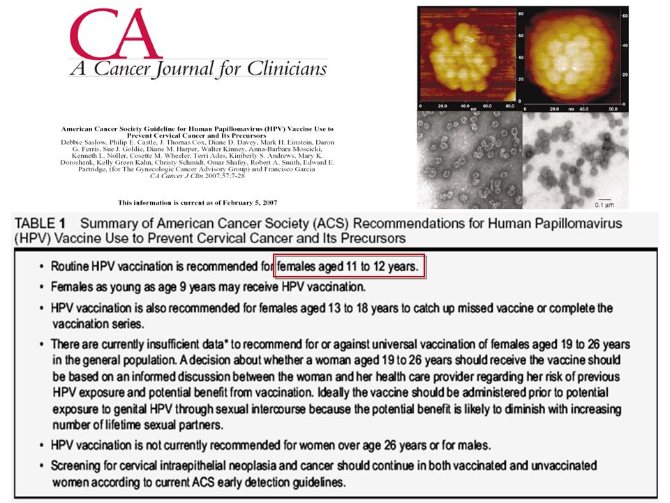
GARDASIL: From Bench Top to Bed-side Vaccine charasteristics non-infectious recombinant (yeast) non-infectious recombinant (yeast) quadrivalent (capsid prt of HPV types 6, 11, 16, 18) quadrivalent (capsid prt of HPV types 6, 11, 16, 18) approved for women aged 9-26 years approved for women aged 9-26 years prevention of cervical cancer & genital wards, prevention of cervical cancer & genital wards, vulvar and vaginal precancerous lesions Vaccine charasteristics non-infectious recombinant (yeast) non-infectious recombinant (yeast) quadrivalent (capsid prt of HPV types 6, 11, 16, 18) quadrivalent (capsid prt of HPV types 6, 11, 16, 18) approved for women aged 9-26 years approved for women aged 9-26 years prevention of cervical cancer & genital wards, prevention of cervical cancer & genital wards, vulvar and vaginal precancerous lesions Clinical trials Phase I : 2,391 women, efficacy 100 % over 40 months Phase I : 2,391 women, efficacy 100 % over 40 months Phase II : 552 women, efficacy 100 % over 5 years Phase II : 552 women, efficacy 100 % over 5 years Phase III : 18,000 women, efficacy 100 % over 2 years Phase III : 18,000 women, efficacy 100 % over 2 years Clinical trials Phase I : 2,391 women, efficacy 100 % over 40 months Phase I : 2,391 women, efficacy 100 % over 40 months Phase II : 552 women, efficacy 100 % over 5 years Phase II : 552 women, efficacy 100 % over 5 years Phase III : 18,000 women, efficacy 100 % over 2 years Phase III : 18,000 women, efficacy 100 % over 2 years
 non-infectious recombinant (yeast) quadrivalent (capsid prt of HPV types 6, 11, 16, 18) quadrivalent (capsid prt of HPV types 6, 11, 16, 18) approved for women aged 9-26 years approved for women aged 9-26 years prevention of cervical cancer & genital wards, prevention of cervical cancer & genital wards, vulvar and vaginal precancerous lesions Vaccine charasteristics non-infectious recombinant (yeast) non-infectious recombinant (yeast) quadrivalent (capsid prt of HPV types 6, 11, 16, 18) quadrivalent (capsid prt of HPV types 6, 11, 16, 18) approved for women aged 9-26 years approved for women aged 9-26 years prevention of cervical cancer & genital wards, prevention of cervical cancer & genital wards, vulvar and vaginal precancerous lesions Clinical trials Phase I : 2,391 women, efficacy 100 % over 40 months Phase I : 2,391 women, efficacy 100 % over 40 months Phase II : 552 women, efficacy 100 % over 5 years Phase II : 552 women, efficacy 100 % over 5 years Phase III : 18,000 women, efficacy 100 % over 2 years Phase III : 18,000 women, efficacy 100 % over 2 years Clinical trials Phase I : 2,391 women, efficacy 100 % over 40 months Phase I : 2,391 women, efficacy 100 % over 40 months Phase II : 552 women, efficacy 100 % over 5 years Phase II : 552 women, efficacy 100 % over 5 years Phase III : 18,000 women, efficacy 100 % over 2 years Phase III : 18,000 women, efficacy 100 % over 2 years")
87
Mechanisms we should concentrate on Increase immune response Enhance cell-mediated immunity Increase tumor-specific immune responses Direct effect on tumor cells Restore immunocompetence Induce prolonged tumor immunity Remove inhibitory factors (eg. tumor antigen immune complexes)
.")
88
To μέλλον των αντικαρκινικών εμβολίων και της ανοσοθεραπείας του καρκίνου Συνέχιση των δοκιμών των εμβολίων σε μικρά φάσης Ι και ΙΙ πρωτόκολλα, χρησιμοποιώντας διάφορα Ag σε διάφορα σχήματα ελπίζοντας στην βελτίωση των κλινικών αποτελεσμάτων πχ. αντι-ιδιοτυπικό εμβόλιο για τα Β-λεμφώματα Αποτέλεσμα: εμβόλια για λίγους ασθενείς σε μεγάλα κέντρα των αναπτυγμένων χωρών Δοκιμή των αποτελεσματικών εμβολίων για την πρόληψη του καρκίνου σε ομάδες υψηλού κινδύνου. Να δοθεί έμφαση στις προκαρκινικές νοσολογικές καταστάσεις. πχ. mucin-1, CEA, HER-2 Αποτέλεσμα: πρόληψη δημιουργίας όγκων πρόληψη δημιουργίας όγκων ευρύτερη εφαρμογή

Παρόμοιες παρουσιάσεις


, Performance Indicators (PIs), Key Performance Indicators (KPIs)>")


 μηρυκαστικά,>")


. Η αντίληψη αποτελεί δημιούργημα του εγκεφάλου.>")